Monitoring response to radiotherapy in human squamous cell cancer bearing nude mice: comparison of 2'-deoxy-2'-[18F]fluoro-D-glucose (FDG) and 3'-[18F]fluoro-3'-deoxythymidine (FLT)
- PMID: 17643202
- PMCID: PMC2040178
- DOI: 10.1007/s11307-007-0104-5
Monitoring response to radiotherapy in human squamous cell cancer bearing nude mice: comparison of 2'-deoxy-2'-[18F]fluoro-D-glucose (FDG) and 3'-[18F]fluoro-3'-deoxythymidine (FLT)
Abstract
Objective: The uptake of 3'-[18F]fluoro-3'-deoxythymidine (FLT), a proliferation marker, was measured before and during fractionated radiotherapy to evaluate the potential of FLT-positron emission tomography (PET) imaging as an indicator of tumor response compared to 2'-deoxy-2'-[18F]fluoro-D-glucose (FDG).
Materials and methods: Nude mice bearing established human head and neck xenografts (HNX-OE; nu/nu mice) were locally irradiated (three fractions/week; 22 Gy) using a 150-kVp unit. Multiple FDG- and FLT-PET scans were acquired during treatment. Tumor volume was determined regularly, and tissue was analyzed for biomarkers involved in tracer uptake.
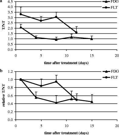
Results: Both groups revealed a significant decline in tumor volume (P<0.01) compared to untreated tumors. For FDG as well as for FLT, a significant decline in retention was observed at day 4. For FLT, most significant decline in retention was observed at day 12; whereas, for FDG, this was already noted at day 4. Maximum decline in tumor-to-nontumor ratios (T/NT) for FDG and FLT was 42+/-18% and 49+/-16% (mean+/-SD), respectively. FLT uptake was higher then that of FDG. For FLT, statistical significant correlations were found for both tumor volume at baseline and at day 29 with T/NT and DeltaT/NT. All tumors demonstrated expression of glucose transporter-1, thymidine kinase-1, and hexokinase II. No differences were found for amount of tumor cells and necrosis at the end of treatment.
Conclusion: This new experimental in vivo model supports the promise of using FLT-PET, as with FDG-PET, to monitor response to external radiotherapy. This warrants further clinical studies to compare these two tracers especially in cancers treated with radiotherapy.
Figures


Similar articles
-
[18F]FLT and [18F]FDG PET for non-invasive treatment monitoring of the nicotinamide phosphoribosyltransferase inhibitor APO866 in human xenografts.PLoS One. 2013;8(1):e53410. doi: 10.1371/journal.pone.0053410. Epub 2013 Jan 4. PLoS One. 2013. PMID: 23308217 Free PMC article.
-
Histopathologic validation of 3'-deoxy-3'-18F-fluorothymidine PET for detecting tumor repopulation during fractionated radiotherapy of human FaDu squamous cell carcinoma in nude mice.Radiother Oncol. 2014 Jun;111(3):475-81. doi: 10.1016/j.radonc.2014.04.002. Epub 2014 May 8. Radiother Oncol. 2014. PMID: 24813091
-
Using dual-tracer PET to predict the biologic behavior of human colorectal cancer.J Nucl Med. 2009 Nov;50(11):1857-64. doi: 10.2967/jnumed.109.064238. Epub 2009 Oct 16. J Nucl Med. 2009. PMID: 19837754
-
Monitoring of anti-cancer treatment with (18)F-FDG and (18)F-FLT PET: a comprehensive review of pre-clinical studies.Am J Nucl Med Mol Imaging. 2015 Oct 12;5(5):431-56. eCollection 2015. Am J Nucl Med Mol Imaging. 2015. PMID: 26550536 Free PMC article. Review.
-
Fludeoxyglucose F18.2023 Oct 15. Drugs and Lactation Database (LactMed®) [Internet]. Bethesda (MD): National Institute of Child Health and Human Development; 2006–. 2023 Oct 15. Drugs and Lactation Database (LactMed®) [Internet]. Bethesda (MD): National Institute of Child Health and Human Development; 2006–. PMID: 30000776 Free Books & Documents. Review.
Cited by
-
[18F]FDG and [18F]FLT positron emission tomography imaging following treatment with belinostat in human ovary cancer xenografts in mice.BMC Cancer. 2013 Apr 1;13:168. doi: 10.1186/1471-2407-13-168. BMC Cancer. 2013. PMID: 23548101 Free PMC article.
-
Early detection of response to experimental chemotherapeutic Top216 with [18F]FLT and [18F]FDG PET in human ovary cancer xenografts in mice.PLoS One. 2010 Sep 24;5(9):e12965. doi: 10.1371/journal.pone.0012965. PLoS One. 2010. PMID: 20885974 Free PMC article.
-
Thymidine Kinase 1 Expression Correlates with Tumor Aggressiveness and Metastatic Potential in OSCC.Diagnostics (Basel). 2025 Jun 19;15(12):1567. doi: 10.3390/diagnostics15121567. Diagnostics (Basel). 2025. PMID: 40564887 Free PMC article.
-
Monitoring early responses to irradiation with dual-tracer micro-PET in dual-tumor bearing mice.World J Gastroenterol. 2010 Nov 21;16(43):5416-23. doi: 10.3748/wjg.v16.i43.5416. World J Gastroenterol. 2010. PMID: 21086558 Free PMC article.
-
Combination Therapy with Trastuzumab and Niraparib: Quantifying Early Proliferative Alterations in HER2+ Breast Cancer Models.Biomedicines. 2023 Jul 25;11(8):2090. doi: 10.3390/biomedicines11082090. Biomedicines. 2023. PMID: 37626587 Free PMC article.
References
-
- None
- Lowe VL SJB (2003) PET imaging head and neck cancer. In: Valk PE, Bailey DL, Townsend DW, Maisey MN (eds) Positron emission tomography. London: Springer
-
- {'text': '', 'ref_index': 1, 'ids': [{'type': 'DOI', 'value': '10.1002/ijc.20379', 'is_inner': False, 'url': 'https://doi.org/10.1002/ijc.20379'}, {'type': 'PubMed', 'value': '15382034', 'is_inner': True, 'url': 'https://pubmed.ncbi.nlm.nih.gov/15382034/'}]}
- Kim MM, Califano JA (2004) Molecular pathology of head-and-neck cancer. Int J Cancer 112:545–553 - PubMed
-
- {'text': '', 'ref_index': 1, 'ids': [{'type': 'DOI', 'value': '10.1001/archotol.130.12.1361', 'is_inner': False, 'url': 'https://doi.org/10.1001/archotol.130.12.1361'}, {'type': 'PubMed', 'value': '15611393', 'is_inner': True, 'url': 'https://pubmed.ncbi.nlm.nih.gov/15611393/'}]}
- Schwartz DL, Rajendran J, Yueh B, et al. (2004) FDG-PET prediction of head and neck squamous cell cancer outcomes. Arch Otolaryngol Head Neck Surg 130:1361–1367 - PubMed
-
- {'text': '', 'ref_index': 1, 'ids': [{'type': 'DOI', 'value': '10.1007/s00405-003-0581-3', 'is_inner': False, 'url': 'https://doi.org/10.1007/s00405-003-0581-3'}, {'type': 'PubMed', 'value': '12736744', 'is_inner': True, 'url': 'https://pubmed.ncbi.nlm.nih.gov/12736744/'}]}
- Hardisson D (2003) Molecular pathogenesis of head and neck squamous cell carcinoma. Eur Arch Otorhinolaryngol 260:502–508 - PubMed
-
- {'text': '', 'ref_index': 1, 'ids': [{'type': 'DOI', 'value': '10.1016/S1536-1632(03)00102-1', 'is_inner': False, 'url': 'https://doi.org/10.1016/s1536-1632(03)00102-1'}, {'type': 'PubMed', 'value': '14499141', 'is_inner': True, 'url': 'https://pubmed.ncbi.nlm.nih.gov/14499141/'}]}
- Klabbers BM, Lammertsma AA, Slotman BJ (2003) The value of positron emission tomography for monitoring response to radiotherapy in head and neck cancer. Mol Imag Biol 5:257–270 - PubMed
Publication types
MeSH terms
Substances
LinkOut - more resources
Full Text Sources
Other Literature Sources
Medical
Research Materials

