. 2007 Jul 20;27(2):197-213.
doi: 10.1016/j.molcel.2007.05.033.

HDAC6 modulates cell motility by altering the acetylation level of cortactin

Affiliations

HDAC6 modulates cell motility by altering the acetylation level of cortactin

Xiaohong Zhang et al. Mol Cell. .

Abstract

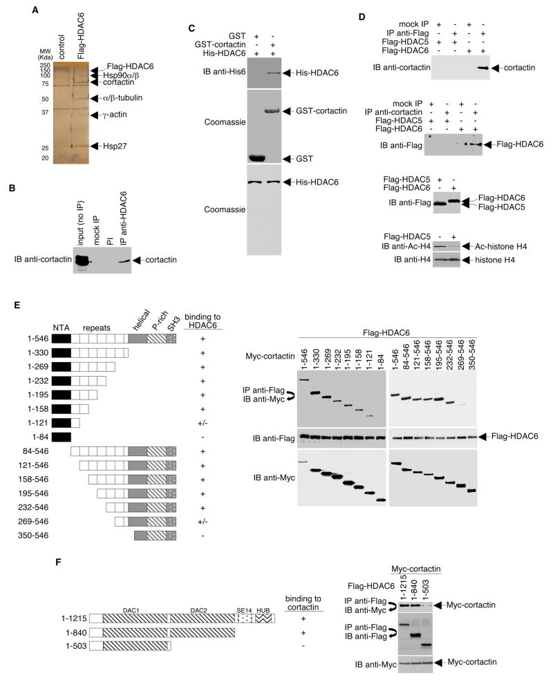
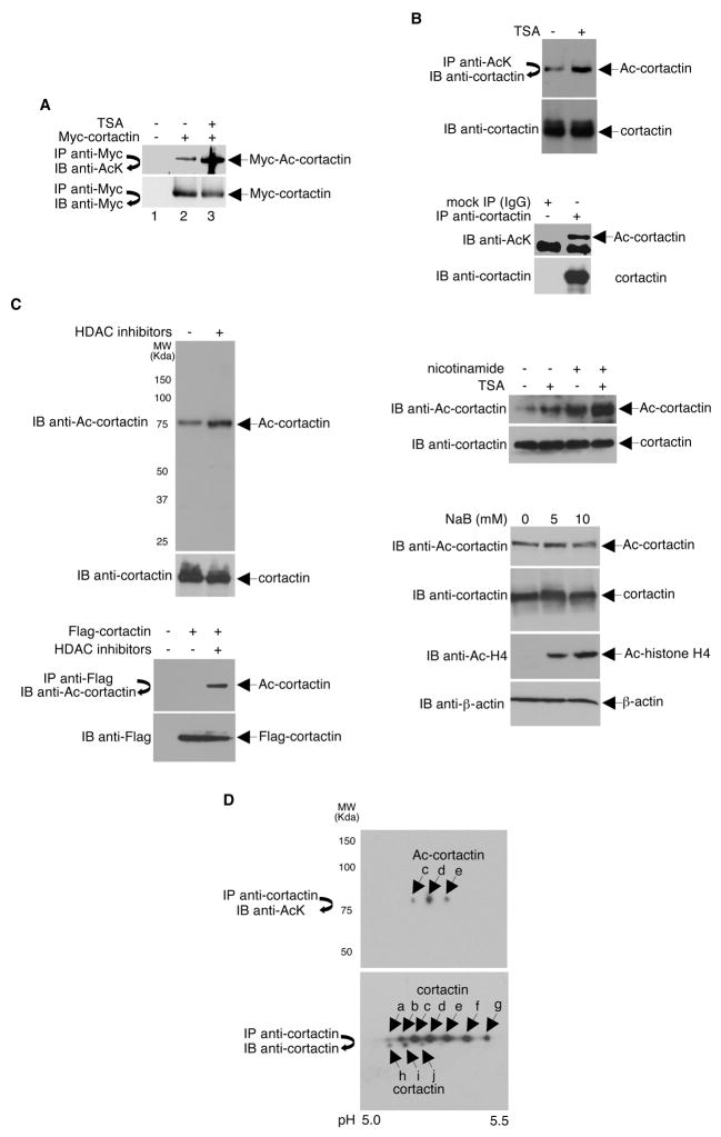
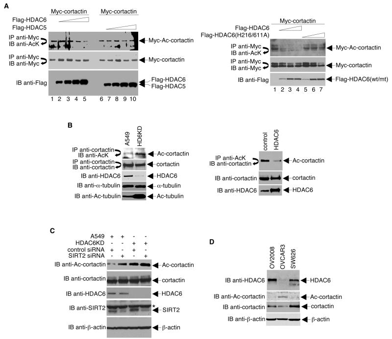
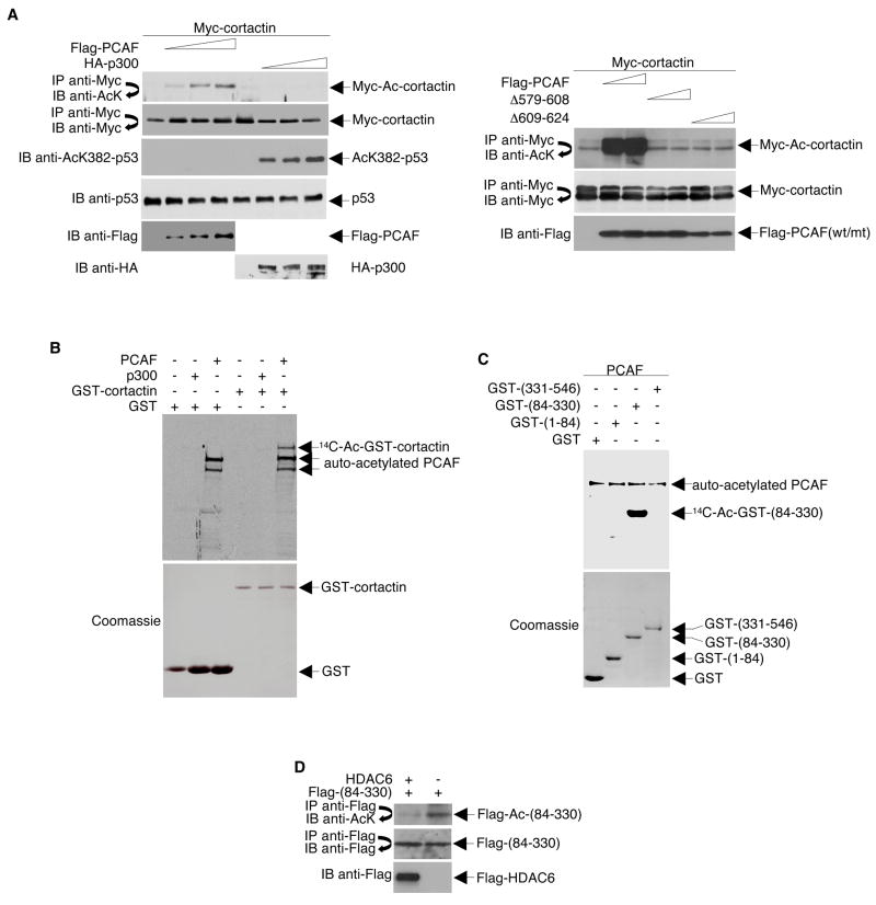
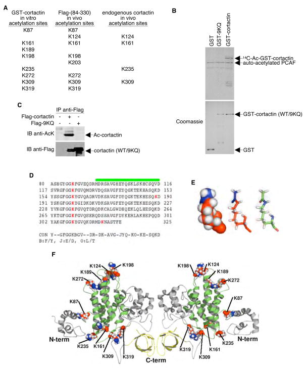
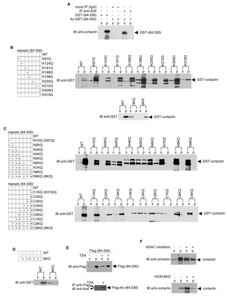
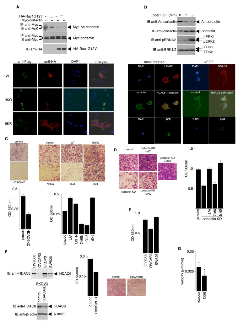
Histone deacetylase 6 (HDAC6) is a tubulin-specific deacetylase that regulates microtubule-dependent cell movement. In this study, we identify the F-actin-binding protein cortactin as a HDAC6 substrate. We demonstrate that HDAC6 binds cortactin and that overexpression of HDAC6 leads to hypoacetylation of cortactin, whereas inhibition of HDAC6 activity leads to cortactin hyperacetylation. HDAC6 alters the ability of cortactin to bind F-actin by modulating a "charge patch" in its repeat region. Introduction of charge-preserving or charge-neutralizing mutations in this cortactin repeat region correlates with the gain or loss of F-actin binding ability, respectively. Cells expressing a charge-neutralizing cortactin mutant were less motile than control cells or cells expressing a charge-preserving mutant. These findings suggest that, in addition to its role in microtubule-dependent cell motility, HDAC6 influences actin-dependent cell motility by altering the acetylation status of cortactin, which, in turn, changes the F-actin binding activity of cortactin.


Figures

Figure 1
Figure 1. HDAC6 Interacts with Cortactin
(A) Silver-stained SDS-PAGE of the immunopurified Flag-tagged HDAC6-containing complexes. “Control” indicates an anti-Flag immunopurified sample prepared from HeLa cells expressing the empty Flag vector. (B) HeLa cell lysate was incubated with protein A-agarose alone (mock IP), preimmune serum (PI), or anti-HDAC6 antibody. Precipitates and total cell lysate (input) were analyzed by Western blotting using an anti-cortactin antibody. (C) GST and GST-cortactin coupled to Sepharose beads were incubated with purified His-HDAC6. The beads were washed extensively, and bound proteins were eluted and analyzed by Western blotting with anti-His6 antibodies (top panel). Coomassie blue stain gels were used to assess the quality and quantities of the purified proteins (middle and bottom panels). (D) HeLa cells were infected with adenoviruses encoding either Flag-tagged HDAC5 or Flag-tagged HDAC6. Whole cell lysates were immunoprecipitated with Flag-specific antibodies and Western blotted with cortactin antibodies (top panel) or vice versa (upper middle panel). Levels of Flag-tagged HDAC5 and Flag-tagged HDAC6 were determined by Western blot analysis of cell extracts using an anti-Flag antibody (lower middle panel). The ability of Flag-HDAC5 to deacetylate histone H4 was assessed by Western blot analysis of cell extracts using an anti-Ac-H4 antibody. (E) Left panel, a schematic diagram (not drawn to scale) of cortactin and various cortactin deletion mutants. P-rich: proline-rich. NTA: N-terminal acidic domain. For simplicity, the Myc portions of the fusion proteins are not shown. Right panel, HeLa cells were infected with adenoviruses encoding Flag-tagged HDAC6 and were transfected with plasmids encoding Myc-tagged cortactin or cortactin deletion mutants. Anti-Flag immunoprecipitates were Western blotted with antibodies specific for Myc (right top panel). Levels of Flag-tagged HDAC6 (right middle panel) and Myc-tagged cortactin (right bottom panel) were determined by Western blot analysis of cell extracts using antibodies specific for Flag or Myc, respectively. To maximize resolution of the different protein fragments, gels shown were prepared with different percentages of polyacrylamide. (F) Left panel, a schematic diagram (not drawn to scale) of HDAC6 and two HDAC6 deletion mutants. DAC: deacetylase (catalytic) domain. SE14: Ser/Glu-containing tetradecapeptide repeats. HUB: HDAC6/USP3/BRAP2-like ubiquitin-binding zinc finger. For simplicity, the Flag portions of the fusion proteins are not shown. Right panel, HeLa cells were transfected with plasmids encoding Myc-tagged cortactin and either Flag-tagged wildtype or mutant HDAC6. Anti-Flag immunoprecipitates were Western blotted with anti-Myc (top panel) or anti-Flag (middle panel). Expression levels of Myc-cortactin were assayed by Western blot with anti-Myc antibodies (bottom panel).
Figure 2
Figure 2. Cortactin is Acetylated in vivo
(A) HeLa cells transfected with either an empty vector or a vector encoding Myc-tagged cortactin were treated with ethanol (−) or 400 ng/ml TSA (+). Anti-Myc immunoprecipitates were Western blotted using antibodies specific for acetyl-lysine (AcK) or Myc. Relative level of acetylated cortactin: Ac-cortactin/cortactin = 0.33 (lane 2), 1.75 (lane 3). (B) HeLa cells were untreated (bottom panel) or treated (top panel) with either ethanol (−) or 400 ng/ml TSA (+) for 12 h. Lysates were immunoprecipitated under high stringency conditions and immunoprecipitates were Western blotted with the indicated antibodies. The same lysates were directly immunoblotted with an anti-cortactin antibody. (C) Left top panel, total extracts prepared from HeLa cells treated with ethanol (−) or 400 ng/ml TSA plus 20 mM nicotinamide (+) for 12 h were separated on SDS-PAGE and analyzed by Western blot with the anti-acetyl-cortactin antibody. The blot was stripped and re-probed with an anti-cortactin antibody. Left bottom panel, HeLa cells transfected with plasmids expressing Flag-tagged cortactin or an empty parental vector were treated with ethanol (−) or 400 ng/ml TSA plus 20 mM nicotinamide (+) for 12 h. Cell lysates were immunoprecipitated with anti-Flag and Western blotted with an anti-acetyl-cortactin antibody or directly Western blotted with a Flag-specific antibody. Right panels, extracts prepared from cells treated with nicotinamide, sodium butyrate (NaB), and/or TSA were analyzed as before. Anti-Ac-H4 blot was performed to confirm that NaB was active in this system. Anti-cortactin and anti-β-actin Western blots were done as loading controls. (D) Top panel, anti-cortactin immunoprecipitates prepared from a NIH3T3 whole cell extract was separated on a 2-dimensional gel and Western blotted with an anti-acetyl-lysine antibody. Bottom panel, the blot was stripped and re-probed with an anti-cortactin antibody.
Figure 3
Figure 3. HDAC6 Deacetylates Cortactin in vivo
(A) Left panel, Myc-tagged cortactin was expressed in HeLa cells by transient transfection. Various amounts of Flag-tagged HDAC6 and Flag-tagged HDAC5 were expressed in HeLa cells by adenoviral infection. Right panel, Myc-cortactin, Flag-HDAC6, and Flag-tagged HDAC6 catalytically defective mutant (H216/611A) were expressed in HeLa cells by transfection. Anti-Myc immunoprecipitates were Western blotted with antibodies specific to either acetyl-lysine (top panels) or Myc (middle panels). Cell extracts were Western blotted with a Flag-specific antibody (bottom panels). Relative level of acetylated cortactin: Ac-cortactin/cortactin = 0.83 (lane 1, left panel), 0.17 (lane 5, left panel), 0.8 (lane 6, left panel), 0.9 (lane 10, left panel), 0.65 (lane 1, right panel), 0.03 (lane 4, right panel), 0.57 (lane 7, right panel). (B) Left panel, lysates prepared from A549 cells and A549 cells stably expressing HDAC6 siRNA (HD6KD) were immunoprecipitated with a cortactin antibody, and the immunoprecipitates were Western blotted with an acetyl-lysine-specific antibody (top) or an anti-cortactin antibody (second from the top). Lysates were also directly Western blotted with antibodies specific for HDAC6, α-tubulin, and acetylated α-tubulin. Right panel, cell lysates were prepared from stably transfected HeLa cells expressing either the empty vector or high levels of HDAC6 and used either in a high stringency immunoprecipitation with anti-acetyl-lysine-specific antibodies followed by Western blotting with anti-cortactin (top) or for Western blotting with antibodies specific for either cortactin or HDAC6 (middle and bottom panels, respectively). (C) Four different sets of SIRT2 siRNA from the siGENOME SMART pool were purchased from Dharmacon and used to knock-down SIRT2 expression in A549 or HDAC6KD cells. Protein expression levels and the effects on cortactin acetylation were examined by Western blotting with the indicated antibodies. * indicates a non-specific band on the anti-SIRT2 Western blot. (D) Cell lysates prepared from three different ovarian cancer cell lines subjected to Western blot analysis with the indicated antibodies.
Figure 4
Figure 4. Cortactin is Acetylated Primarily in its Repeat Region
(A) HeLa cells were transfected with Myc-tagged cortactin and various amounts of Flag-tagged PCAF, Flag-tagged PCAF catalytically-dead mutants, or HA-tagged p300. Anti-Myc immunoprecipitates were Western blotted with antibodies specific for either acetyl-lysine or Myc. Levels of Flag-tagged PCAF and HA-tagged p300 were determined by Western blotting of cell extracts with antibodies specific for Flag and HA, respectively. To confirm that p300 was active in this system, a Western blot was performed using the same extracts with an antibody specific for acetylated K382 of p53. (B) GST-cortactin was incubated with 14C-acetyl CoA and recombinant PCAF or p300. Acetylation of GST-cortactin was visualized by autoradiography of SDS-PAGE gels (top panel). Levels of GST-cortactin were determined by Coomassie staining of SDS-PAGE gels (bottom panel). (C) Acetylation assays were performed using purified GST-tagged cortactin fragments and PCAF. (D) HeLa cells were infected with adenoviruses encoding either GFP (−) or Flag-tagged HDAC6 (+). Whole cell lysates were immunoprecipitated with Flag-specific antibodies and Western blotted with antibodies against acetylated lysines (top panel). The blot was stripped and re-probed with anti-Flag (middle panel). A direct Western blot was performed to assess expression of Flag-HDAC6 (bottom panel).
Figure 5
Figure 5. Identification of Acetylation Sites of Cortactin in vitro and in vivo
(A) GST-tagged cortactin (in vitro acetylated by PCAF), Flag-tagged cortactin (expressed and purified from HeLa cells), and endogenous cortactin immunopurified with anti-cortactin antibody were cleaved with trypsin and analyzed by ion trap mass spectrometry. Positions of unambiguously identified acetylated lysine residues are listed. (B) In vitro acetylation assays performed using the catalytic domain of PCAF and GST-tagged (wildtype) or 9KQ mutant cortactin. Reaction products were resolved by SDS-PAGE. Acetylated proteins were visualized by autoradiography (top panel), and total amounts of protein were monitored by Coomassie staining (bottom panel). (C) Flag-tagged cortactin (wildtype) and Flag-tagged 9KQ mutant cortactin were expressed in HeLa cells by transient transfection. Anti-Flag immunoprecipitates were Western blotted with antibodies specific to either acetyl-lysine or Flag. (D) Alignment of the mouse cortactin repeats (residues 80 to 325). The green line represents the predicted transmembrane zone, and the red bold letters represent the potential acetylated lysine residues. (E) Representation of the acetylated residues in balls (left) and sticks (center and right). Atoms in red belong to the lysine residues and atoms in blue to the acetyl groups. The molecule on the right is colored according to atom type: carbon (green), nitrogen (blue), oxygen (red), and hydrogen (white). (F) Cartoon representations of both sides of the mouse cortactin model. The cortactin repeats are in green and the SH3 motif in the carboxyl-terminal is in yellow. The acetylated lysines are in the same color scheme described in E (left and center).
Figure 6
Figure 6. Acetylation of Cortactin Reduces its Interaction with F-actin
(A) GST-tagged cortactin (the repeat region) was acetylated by PCAF in vitro. Reaction mixtures were then incubated with mouse IgG or anti-acetyl-lysine antibodies. Precipitates were resolved by SDS-PAGE. The band corresponding to the repeat region was eluted from the gel with 1% SDS. Eluates were incubated with F-actin, and co-sedimentation assays were performed. Supernatants (S) and pellets (P) were subjected to Western blotting with cortactin-specific antibodies. For lanes 1 and 2, co-sedimentation assays were performed using unacetylated GST-tagged cortactin (the repeat region). (B, C, D) GST-tagged wildtype or mutant cortactin proteins were incubated with F-actin, and co-sedimentation assays were performed. Supernatants and pellets were subjected to Western blotting with an anti-GST antibody. Diagrams of the wildtype and mutant constructs are shown. (E) HeLa cells were transfected with a plasmid encoding Flag-tagged cortactin (repeat region) and treated with ethanol (vehicle control) or 400 ng/ml TSA for 12 h. Top panel, co-sedimentation assays were performed on cell lysates, and supernatants and pellets were subjected to Western blotting with an anti-Flag antibody. Bottom panel, Anti-Flag immunoprecipitates were Western blotted with an antibody specific for acetyl-lysine. (F) Similar co-sedimentation experiments were performed to assess endogenous cortactin-F-actin interactions.
Figure 7
Figure 7. Acetylation of Cortactin Prevents its Localization to Membrane Ruffles and Inhibits Cell Motility
(A) Top panel, NIH3T3 cells were transfected with plasmids that express Myc-cortactin and various amounts of HA-Rac1G12V. Anti-Myc immunoprecipitates prepared from these cells were assayed for cortactin acetylation by Western blotting with an anti-acetyl-lysine antibody. Immunoprecipitation efficiency and HA-Rac1G12V expression were monitored by Western blotting with with anti-Myc and anti-HA, respectively. Bottom panel, NIH 3T3 cells were transfected with plasmids encoding active Rac1 (HA-Rac1G12V) and either Flag-tagged wildtype, Flag-tagged 9KQ mutant, or Flag-tagged 9KR mutant cortactin. Twenty-four hours post-transfection, cells were immunostained with antibodies or stained with 4′,6-diamidino-2-phenyl-indole and analyzed under a confocal microscope. (B) Top panel, NIH3T3 cells were serum-starved overnight and then treated with 20 ng/ml EGF. Lysates were analyzed by Western blotting with anti-acetyl-cortactin antibodies. The blot was stripped and re-probed with anti-cortactin antibodies. For controls, anti-phospho-ERK1/2 and anti-ERK1/2 Western blots were performed to monitor the efficiency of EGF treatment. Bottom panels, NIH3T3 cells were serum-starved overnight and then either mock-treated or treated with 10 ng/ml EGF for 10 min. Endogenous HDAC6 and cortactin were detected using anti-HDAC6 and anti-cortactin specific antibodies and Alexa-594- and Alexa-488-conjugated secondary antibodies. Flag-9KR and Flag-9KQ were detected with anti-Flag antibodies. (C) Left panels, 293T cells stably expressing HDAC6 siRNA (HDAC6KD; generated by the OligoEngine retrovirus-mediated pSuper RNAi system) and 293T cells expressing control siRNA were serum-starved overnight and assayed for migratory properties. Migratory cells were stained (top) and quantified at OD 560 nm following extraction (bottom). Right panels, parental NIH 3T3 cells (control) and NIH 3T3 cells stably expressing either wildtype or cortactin mutants were serum-starved overnight and assayed for migratory properties as in left panels. (D) Plasmids expressing wildtype or mutant cortactins were transfected into HT1080 cells depleted of cortactin (cortactin KD). Stable polyclonal cell populations were selected and assayed for cell migration activity as in (C). Control cells indicate parental untransfected HT1080 cells. (E) Three different ovarian cancer cell lines were assayed for cell migration activity as in (C). (F) Top left panel, Western blot analysis of HDAC6 expression in different ovarian cancer cell lines. Bottom left panel, Western blots to assess HDAC6 knock-down in SKOV3 cells. Middle and right panels, SKOV3 cells transfected with either HDAC6 siRNA or control siRNA were assayed for cell migration activity as in (C). (G) Data showing velocity of parental MDA-MB-231 cells (control) and a pool of stable cell clones overexpressing 9KQ mutant cortactin undergoing random motility on tissue-culture dishes. For MDA-MB-231 cells, n = 64; for 9KQ stable clones, n = 76. *p < 0.0001 vs. MDA-MB-231 cells. Error bars denote the standard deviation (SD).

Comment in

  • Sirt1 and cell migration.
    Tang BL. Tang BL. Cell Adh Migr. 2010 Apr-Jun;4(2):163-5. doi: 10.4161/cam.4.2.10972. Epub 2010 Apr 18. Cell Adh Migr. 2010. PMID: 20179424 Free PMC article.

References

    1. Bali P, Pranpat M, Bradner J, Balasis M, Fiskus W, Guo F, Rocha K, Kumaraswamy S, Boyapalle S, Atadja P, et al. Inhibition of histone deacetylase 6 acetylates and disrupts the chaperone function of heat shock protein 90: a novel basis for antileukemia activity of histone deacetylase inhibitors. J Biol Chem. 2005;280:26729–26734. - PubMed
    1. Bowden ET, Barth M, Thomas D, Glazer RI, Mueller SC. An invasion-related complex of cortactin, paxillin and PKCmu associates with invadopodia at sites of extracellular matrix degradation. Oncogene. 1999;18:4440–4449. - PubMed
    1. Bowden ET, Onikoyi E, Slack R, Myoui A, Yoneda T, Yamada KM, Mueller SC. Co-localization of cortactin and phosphotyrosine identifies active invadopodia in human breast cancer cells. Exp Cell Res. 2006;312:1240–1253. - PubMed
    1. Boyault C, Gilquin B, Zhang Y, Rybin V, Garman E, Meyer-Klaucke W, Matthias P, Muller CW, Khochbin S. HDAC6-p97/VCP controlled polyubiquitin chain turnover. EMBO J. 2006;25:3357–3366. - PMC - PubMed
    1. Bryce NS, Clark ES, Leysath JL, Currie JD, Webb DJ, Weaver AM. Cortactin promotes cell motility by enhancing lamellipodial persistence. Curr Biol. 2005;15:1276–1285. - PubMed

Publication types

MeSH terms

Associated data

LinkOut - more resources