. 2007 Nov 14;573(1-3):29-38.
doi: 10.1016/j.ejphar.2007.06.049. Epub 2007 Jul 4.

Histone H3 phosphorylation at serine 10 and serine 28 is mediated by p38 MAPK in rat hepatocytes exposed to ethanol and acetaldehyde

Affiliations

Histone H3 phosphorylation at serine 10 and serine 28 is mediated by p38 MAPK in rat hepatocytes exposed to ethanol and acetaldehyde

Youn Ju Lee et al. Eur J Pharmacol. .

Abstract

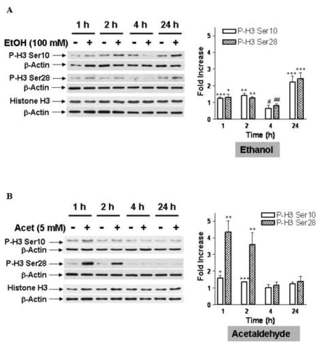
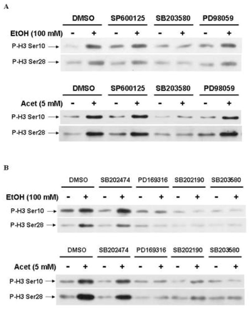
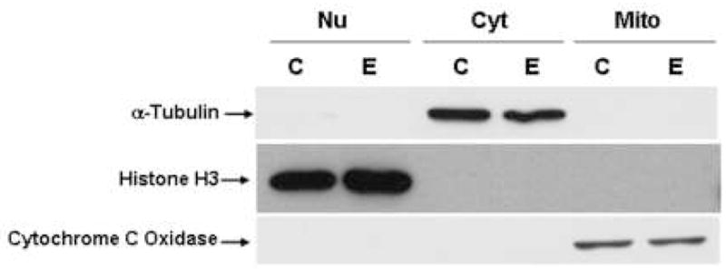
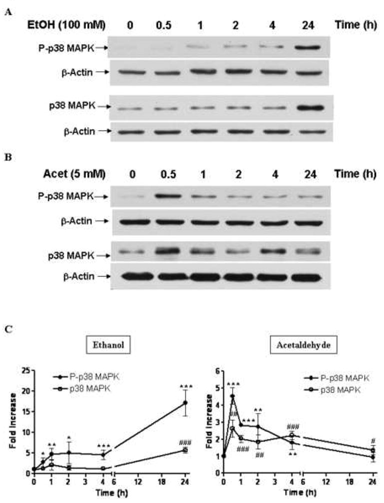
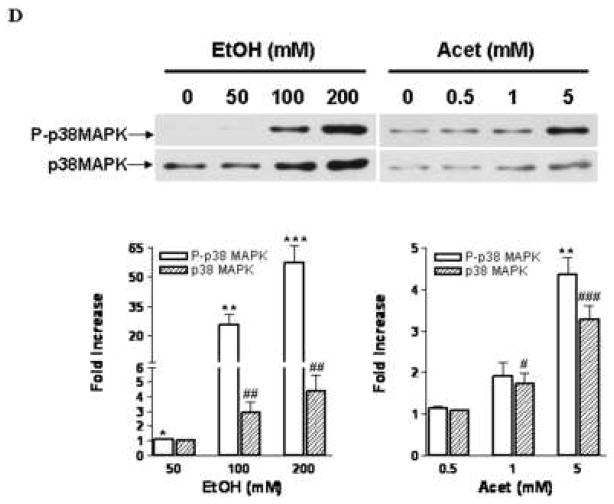
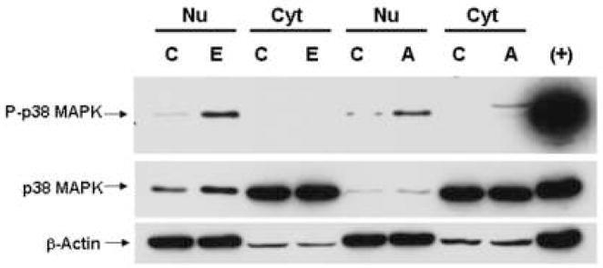
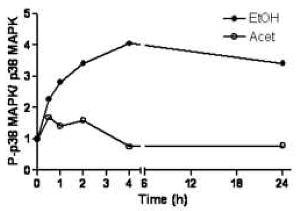
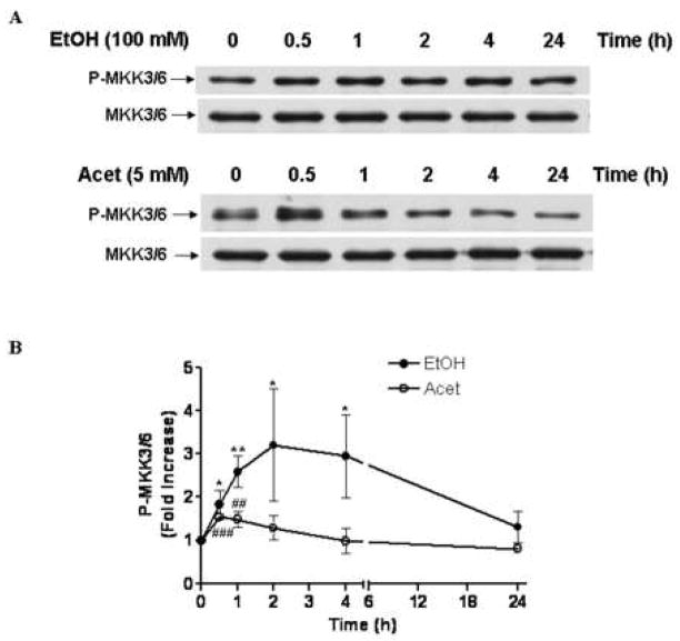
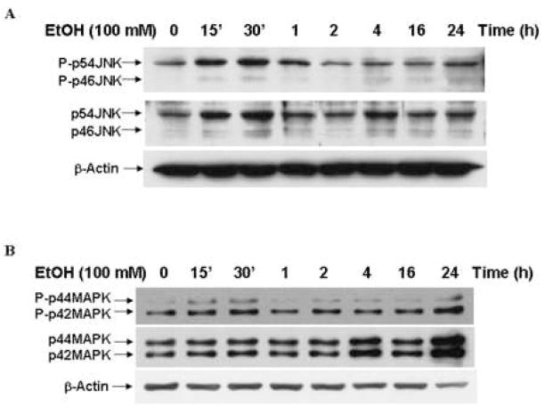
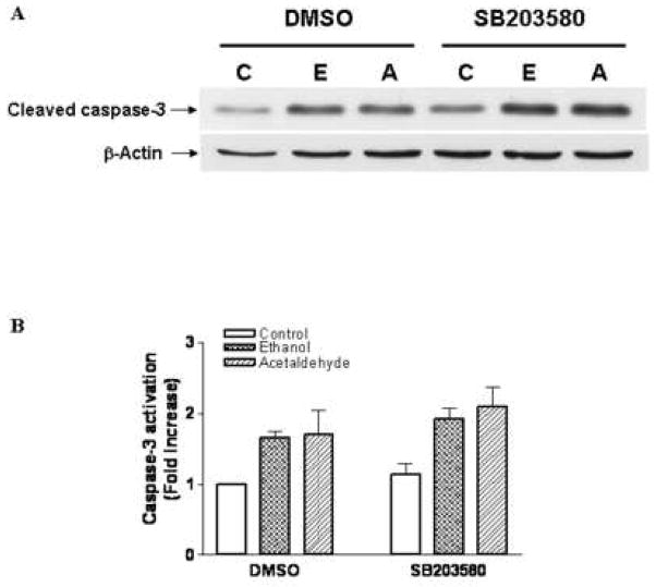
Ethanol modulates mitogen-activated protein kinases (MAPKs). We have now investigated the influence of ethanol and its metabolite, acetaldehyde on histone H3 phosphorylation to ascertain downstream targets of MAPKs. In primary culture of rat hepatocytes, ethanol and acetaldehyde increased phosphorylation of nuclear histone H3 at serine 10 and serine 28. Specific inhibitors of p38 MAPK, SB203580, PD169316 and SB202190 blocked this phosphorylation. The inactive analogue, SB202474 had no effect. In contrast, c-Jun N-terminal kinase (JNK) inhibitor, SP600125 or MAP/ERK kinase (MEK) 1/2 inhibitor, PD98059 had no effect on the histone H3 phosphorylation. The p38 MAPK activation correlated with upstream activation of MAPK kinase (MKK) 3/6 but was independent of protein synthesis. In the nuclear fraction, the phosphorylation of p38 MAPK and its protein level increased with peak activation at 24 h by ethanol and at 30 min by acetaldehyde. These responses were ethanol and acetaldehyde dose dependent. Surprisingly, the phosphorylation of p38 MAPK was undetectable in the cytosolic fraction suggesting a subcellular selectivity of p38 MAPK signaling. The phosphorylation of JNK and p42/44 MAPK and their protein levels also increased in the nuclear fraction. Although ethanol caused translocation of all three major MAPKs (p42/44 MAPK, JNK, p38 MAPK) into the nucleus, histone H3 phosphorylation at serine 10 and serine 28 was mediated by p38 MAPK. This histone H3 phosphorylation had no influence on ethanol and acetaldehyde induced apoptosis. These studies demonstrate for the first time that ethanol and acetaldehyde stimulated phosphorylation of histone H3 at serine 10 and serine 28 are downstream nuclear response mediated by p38 MAPK in hepatocytes.


Figures

Fig. 1
Fig. 1. Phosphorylation of histone H3 at serine 10 and serine 28 by ethanol and acetaldehyde
Hepatocytes were treated with 100 mM ethanol (EtOH) (A) or 5 mM acetaldehyde (Acet) (B) for indicated time. Acid soluble proteins were extracted from nuclear fraction. Western blotting was carried out to detect phospho-histone H3 at serine 10 (P-H3 Ser10), phospho-histone H3 at serine 28 (P-H3 Ser28) and histone H3. The membranes were reprobed with anti-β-actin antibody. Quantitative analysis of bands was performed by Quantity One software (Bio-Rad) which gave intensity values. The ratio of the intensity of P-H3 Ser10 or 28 to β-actin for control and treated samples were determined. The fold increase in ratio over control for each time point is presented in bar graph, where control value represents 1. Values are mean ± S.E.M. (bars), n = 3. *P < 0.05, **P < 0.01, and ***P < 0.001, greater than control; # P < 0.05 and ## P < 0.01, less than control.
Fig. 2
Fig. 2. The effects of inhibitors of MAPKs on phosphorylation of histone H3 at serine 10 and serine 28 by ethanol and acetaldehyde
(A) Hepatocytes were pretreated with DMSO (vehicle control), 20 μM SP600125, 10 μM SB203580, 50 μM PD98059 and then treated with 100 mM ethanol (EtOH) or 5 mM acetaldehyde (Acet) for 24 h or 1 h, respectively. The phosphorylation of histone H3 at serine 10 (P-H3 Ser10) and serine 28 (P-H3 Ser28) were monitored by Western blotting. (B) Hepatocytes were pretreated with DMSO (vehicle control), 10 μM SB202474 (an inactive analogue of p38 MAPK inhibitiors), 10 μM PD169316, 10 μM SB202190, 10 μM SB203580 and then treated with 100 mM ethanol (EtOH) or 5 mM acetaldehyde (Acet) for 24 h or 1 h, respectively. The phosphorylation of histone H3 at serine 10 (P-H3 Ser10) and serine 28 (P-H3 Ser28) were monitored by Western blotting. The data are representative of three independent experiments.
Fig. 3
Fig. 3. Subcellular fractionation
Hepatocytes were treated with 100 mM ethanol for 24 h and then nuclear, cytosolic, and mitochondrial proteins were prepared. Same amount of proteins (30 μg) from each fraction were used for Western blot analysis using antibodies against α-tubulin, histone H3 or cytochrome C oxidase. The data are representative of three independent experiments (C, control; E, ethanol; Nu, nucleus; Cyt, cytosol; Mito, mitochondria).
Fig. 4
Fig. 4. Nuclear translocation of p38 MAPK by ethanol and acetaldehyde
Hepatocytes were treated with 100 mM ethanol (EtOH) (A) or 5 mM acetaldehyde (Acet) (B) for indicated time. Nuclear protein was subjected to Western blot to detect phospho-p38 MAPK (P-p38 MAPK) and p38 MAPK protein. The membranes were reprobed with anti β-actin antibody. The data are representative of three independent experiments. (C) The fold increase in P-p38 MAPK and p38 MAPK were quantitated by Quantity One software (Bio-Rad), where control value represents 1. Values represented are mean ± S.E.M. (bars), n = 3. *P < 0.05, **P < 0.01, and *** P < 0.001 compared with control (time = 0) of P-p38 MAPK; # P < 0.05, ## P < 0.01, and ### P < 0.001 compared with control (time = 0) of p38 MAPK. (D) Hepatocytes were treated with different concentration of ethanol (EtOH) or acetaldehyde (Acet) for 24 h and 30 min, respectively. Nuclear protein was subjected to Western blot analysis to detect phospho-p38 MAPK (P-p38 MAPK) and p38 MAPK protein. The data are representative of three independent experiments. Values represented are mean ± S.E.M. (bars), n = 3. *P < 0.05, **P < 0.01, and *** P < 0.001 compared with control of P-p38 MAPK; # P < 0.05, ## P < 0.01, and ### P < 0.001 compared with control of p38 MAPK.
Fig. 5
Fig. 5. Selective activation of p38 MAPK by ethanol and acetaldehyde in nucleus
Hepatocytes were treated with 100 mM ethanol for 24 h or 5 mM acetaldehyde for 30 min. Nuclear and cytosolic proteins were prepared and same amount of proteins (30 μg) were used for Western blot using anti- phospho-p38 MAPK antibody. The cell extract from C6 cell treated with UV was used as a positive control (+). The membrane was reprobed with anti-p38 MAPK antibody and then reprobed with anti β-actin antibody. The data are representative of three independent experiments (C, control; E, ethanol; A, acetaldehyde; Nu, nucleus; Cyt, cytosol).
Fig. 6
Fig. 6. Ratio of phospho-p38 MAPK (P-p38 MAPK) to p38 MAPK in hepatocyte nucleus after ethanol or acetaldehyde treatment
Ratio of the fold increase in P-p38 MAPK to p38 MAPK after ethanol (EtOH) or acetaldehyde (Acet) for different times is presented.
Fig. 7
Fig. 7. Nuclear activation of MKK3/6 by ethanol and acetaldehyde
(A) Hepatocytes were treated with 100 mM ethanol (EtOH) or 5 mM acetaldehyde (Acet) for indicated time. Nuclear protein was subjected to Western blot to detect phospho-MKK3/6 (P-MKK3/6) and MKK3/6 protein. (B) The fold increase in P-MKK3/6 was quantitated by Quantity One software (Bio-Rad), where control value represents 1. Values represented are mean ± S.E.M. (bars), n = 3. *P < 0.05 and **P < 0.01 compared with control (time=0) by ethanol; # P < 0.05, ## P < 0.01, and ### P < 0.001 compared with control (time=0) by acetaldehyde.
Fig. 8
Fig. 8. Nuclear translocation of JNK and p42/44 MAPK by ethanol
Hepatocytes were treated with 100 mM ethanol (EtOH) for indicated time (0 – 24 h) and nuclear proteins were prepared. The phospho-JNK (P-p46/54 JNK), JNK protein (p46/54 JNK) (A), phospho-p42/44 MAPK (P-p42/44 MAPK) and p42/44 MAPK protein (p42/44 MAPK) (B) were detected by Western Blot. The data are representative of three independent experiments.
Fig. 9
Fig. 9. The effect of SB203580 on caspase-3 activation by ethanol and acetaldehyde
Hepatocytes were pretreated with DMSO (vehicle control) or 10 μM SB203580 for 1 h and then treated with 100 mM ethanol or 5 mM acetaldehyde for 2 h. (A) The activation of caspase-3 was determined by Western blotting with anti-cleaved caspase-3 antibody. (B) The fold increase in caspase-3 activation was quantitated by Quantity One software (Bio-Rad). The data are representative of four independent experiments. Values represented are mean ± S.E.M. (bars), n = 6 (C, control; E, ethanol; A, acetaldehyde).

Similar articles

Cited by

References

    1. Ahn NG, Seger R, Bratlien RL, Diltz CD, Tonks NK, Krebs EG. Multiple components in an epidermal growth factor-stimulated protein kinase cascade. In vitro activation of a myelin basic protein/microtubule-associated protein 2 kinase. J Biol Chem. 1991;266:4220–4227. - PubMed
    1. Aroor AR, Shukla SD. MAP kinase signaling in diverse effects of ethanol. Life Sci. 2004;74:2339–2364. - PubMed
    1. Bennett BL, Sasaki DT, Murray BW, O’Leary EC, Sakata ST, Xu W, Leisten JC, Motiwala A, Pierce S, Satoh Y, Bhagwat SS, Manning AM, Anderson DW. SP600125, an anthrapyrazolone inhibitor of Jun N-terminal kinase. Proc Natl Acad Sci USA. 2001;98:13681–13686. - PMC - PubMed
    1. Cavigelli M, Dolfi F, Claret FX, Karin M. Induction of c-fos expression through JNK-mediated TCF/Elk-1 phosphorylation. EMBO J. 1995;14:5957–5964. - PMC - PubMed
    1. Chadee DN, Hendzel MJ, Tylipski CP, Allis CD, Bazett-Jones DP, Wright JM, Davie JR. Increased Ser-10 phosphorylation of histone H3 in mitogen-stimulated and oncogene-transformed Mouse Fibroblasts. J Biol Chem. 1999;274:24914–24920. - PubMed

Publication types

MeSH terms