

Chicken single-chain variable fragments against the SARS-CoV spike protein
- PMID: 17643500
- PMCID: PMC7112778
- DOI: 10.1016/j.jviromet.2007.06.010
Chicken single-chain variable fragments against the SARS-CoV spike protein
Abstract
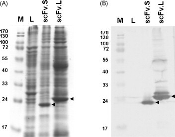
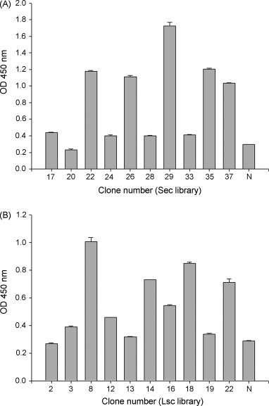
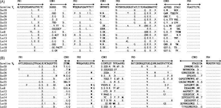
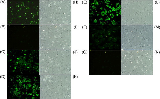
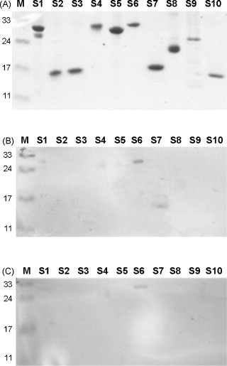
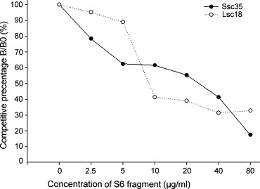
The major concern for severe acute respiratory syndrome (SARS), caused by the SARS-associated coronavirus (SARS-CoV), is the lack of diagnostic and therapeutic agents. Using a phage display technology in a chicken system, high-affinity monoclonal antibody fragments against the SARS-CoV spike protein were characterized. Ten truncated spike protein gene fragments were expressed in Escherichia coli cells. Following the immunization of chickens with these recombinant spike proteins, two single-chain variable fragment (scFv) antibody libraries were established with short or long linkers to contain 5x10(7) and 9x10(6) transformants, respectively. After four rounds of panning selection, the scFv antibodies of randomly chosen clones were demonstrated by Coomassie blue staining, and verified by western blot analysis. In a comparison of nucleotide sequences with the chicken germline gene, we found that all clones varied in the complementarity-determining regions, that two scFv antibodies reacted significantly with SARS-CoV-infected Vero cells, and that those two specific scFv antibodies recognized the same region of the spike protein spanning amino acid residues 750-1000. In conclusion, the results suggest that the chicken scFv phage display system can be a potential model for mass production of high-affinity antibodies against the SARS-CoV spike protein.
Figures
Similar articles
-
A dominant antigenic epitope on SARS-CoV spike protein identified by an avian single-chain variable fragment (scFv)-expressing phage.Vet Immunol Immunopathol. 2007 May 15;117(1-2):75-85. doi: 10.1016/j.vetimm.2007.02.001. Epub 2007 Feb 12. Vet Immunol Immunopathol. 2007. PMID: 17360045 Free PMC article.
-
Human monoclonal antibody combination against SARS coronavirus: synergy and coverage of escape mutants.PLoS Med. 2006 Jul;3(7):e237. doi: 10.1371/journal.pmed.0030237. PLoS Med. 2006. PMID: 16796401 Free PMC article.
-
Molecular and biological characterization of human monoclonal antibodies binding to the spike and nucleocapsid proteins of severe acute respiratory syndrome coronavirus.J Virol. 2005 Feb;79(3):1635-44. doi: 10.1128/JVI.79.3.1635-1644.2005. J Virol. 2005. PMID: 15650189 Free PMC article.
-
Severe acute respiratory syndrome (SARS): development of diagnostics and antivirals.Ann N Y Acad Sci. 2006 May;1067(1):500-5. doi: 10.1196/annals.1354.072. Ann N Y Acad Sci. 2006. PMID: 16804033 Free PMC article. Review.
-
Neutralizing human monoclonal antibodies to severe acute respiratory syndrome coronavirus: target, mechanism of action, and therapeutic potential.Rev Med Virol. 2012 Jan;22(1):2-17. doi: 10.1002/rmv.706. Epub 2011 Sep 8. Rev Med Virol. 2012. PMID: 21905149 Free PMC article. Review.
Cited by
-
Infectious disease antibodies for biomedical applications: A mini review of immune antibody phage library repertoire.Int J Biol Macromol. 2020 Nov 15;163:640-648. doi: 10.1016/j.ijbiomac.2020.06.268. Epub 2020 Jul 8. Int J Biol Macromol. 2020. PMID: 32650013 Free PMC article. Review.
-
An approach towards development of monoclonal IgY antibodies against SARS CoV-2 spike protein (S) using phage display method: A review.Int Immunopharmacol. 2020 Aug;85:106654. doi: 10.1016/j.intimp.2020.106654. Epub 2020 Jun 3. Int Immunopharmacol. 2020. PMID: 32512271 Free PMC article. Review.
-
Antibody-based sensors: principles, problems and potential for detection of pathogens and associated toxins.Sensors (Basel). 2009;9(6):4407-45. doi: 10.3390/s90604407. Epub 2009 Jun 5. Sensors (Basel). 2009. PMID: 22408533 Free PMC article.
-
Recombinant antibodies for diagnostics and therapy against pathogens and toxins generated by phage display.Proteomics Clin Appl. 2016 Oct;10(9-10):922-948. doi: 10.1002/prca.201600002. Epub 2016 Jun 21. Proteomics Clin Appl. 2016. PMID: 27198131 Free PMC article. Review.
-
Phage Display Technique as a Tool for Diagnosis and Antibody Selection for Coronaviruses.Curr Microbiol. 2021 Apr;78(4):1124-1134. doi: 10.1007/s00284-021-02398-9. Epub 2021 Mar 9. Curr Microbiol. 2021. PMID: 33687511 Free PMC article. Review.
References
-
- Abouzid K., Ndeboko B., Durantel S., Jamard C., Zoulim F., Buronfosse T., Cova L. Genetic vaccination for production of DNA-designed antibodies specific to Hepadnavirus envelope proteins. Vaccine. 2006;24:4615–4617. - PubMed
-
- Andris-Widhopf J., Rader C., Steinberger P., Fuller R., Barbas C.F., III Methods for the generation of chicken monoclonal antibody fragments by phage display. J. Immunol. Methods. 2000;242:159–181. - PubMed
-
- Barbas C.F., III, Burton D.R., Scott J.K., Silverman G.J. Cold Spring Harbor Laboratory Press; Cold Spring Harbor, NY: 2001. Phage Display: A Laboratory Manual.
-
- Bird R.E., Hardman K.D., Jacobson J.W., Johnson S., Kaufman B.M., Lee S.M., Lee T., Pope S.H., Riordan G.S., Whitlow M. Single-chain antigen-binding proteins. Science. 1988;242:423–426. - PubMed
Publication types
MeSH terms
Substances
LinkOut - more resources
Full Text Sources
Miscellaneous
