Review
. 2007 Aug;17(4):264-71.
doi: 10.1016/j.gde.2007.05.010. Epub 2007 Jul 23.

Understanding morphogen gradients: a problem of dispersion and containment

Affiliations
Review

Understanding morphogen gradients: a problem of dispersion and containment

Thomas B Kornberg et al. Curr Opin Genet Dev. 2007 Aug.

Abstract

Protein morphogens are instructive signals that regulate growth and patterning of tissues and organs. They form long-range, dynamic gradients by moving from regions of high concentration (producing cells) to regions of low concentration (the adjacent, nonproducing developmental field). Since morphogen activity must be limited to the adjacent target field, we want to understand both how signaling proteins move and how their dispersion is restricted. We consider the variety of settings for long-range morphogen systems in Drosophila. In the early embryo, morphogens appear to disperse by free diffusion, and impermeable membranes physically constrain them. However, at later stages, containment is achieved without physical barriers. We argue that in the absence of constraining barriers, gradient-generating dispersion of morphogens cannot be achieved by passive diffusion and that other mechanisms for distribution must be considered.


Figures

Figure 1
Figure 1
Morphogen gradient systems in the Drosophila oocyte and early embryo. (A) A/P & D/V Axes (Oocyte) - Definition of the anteroposterior and dorsoventral axes of the embryo initiates during oogenesis when gurken is expressed by the posteriorly situated oocyte nucleus (orange) at stage 6-7. At this stage, the oocyte is relatively small and is juxtaposed to nurse cells (brown) and to somatic follicle cells (gray). All follicle cells express the EGFR Gurken receptor (purple), but the posterior follicle cells closest to oocyte nucleus presumably receive most of the secreted Gurken protein (green) and are activated (yellow). At stage 10, the oocyte nucleus has assumed an anterodorsal position, and upon expression of gurken, EGFR activation induces dorsal cell fates among the nearby follicle cells. (B) A/P Axis (Embryo) – Post-fertilization, bicoid (blue) RNA and nanos RNA (red) sequestered at the A and P poles, respectively, are translated; Bicoid and Nanos proteins disperse across the syncytium. (C) Terminal system – Inactive pro-Trunk (lime green) and the Torso receptor (purple) are distributed uniformly in the perivitelline fluid and embryo plasma membrane, respectively. Following proteolytic activation initiated by follicle cells at the A and P poles, active Trunk (dark green) disperses and activates Torso (yellow). (D) D/V Axis – Inactive pro-Spaetzle (burgundy) and the Toll (purple) are distributed uniformly in the perivitelline fluid and embryo plasma membrane, respectively. Following proteolytic activation initiated by follicle cells along the ventral midline, active Spaetzle (red) disperses and activates Toll (yellow).
Figure 1
Figure 1
Morphogen gradient systems in the Drosophila oocyte and early embryo. (A) A/P & D/V Axes (Oocyte) - Definition of the anteroposterior and dorsoventral axes of the embryo initiates during oogenesis when gurken is expressed by the posteriorly situated oocyte nucleus (orange) at stage 6-7. At this stage, the oocyte is relatively small and is juxtaposed to nurse cells (brown) and to somatic follicle cells (gray). All follicle cells express the EGFR Gurken receptor (purple), but the posterior follicle cells closest to oocyte nucleus presumably receive most of the secreted Gurken protein (green) and are activated (yellow). At stage 10, the oocyte nucleus has assumed an anterodorsal position, and upon expression of gurken, EGFR activation induces dorsal cell fates among the nearby follicle cells. (B) A/P Axis (Embryo) – Post-fertilization, bicoid (blue) RNA and nanos RNA (red) sequestered at the A and P poles, respectively, are translated; Bicoid and Nanos proteins disperse across the syncytium. (C) Terminal system – Inactive pro-Trunk (lime green) and the Torso receptor (purple) are distributed uniformly in the perivitelline fluid and embryo plasma membrane, respectively. Following proteolytic activation initiated by follicle cells at the A and P poles, active Trunk (dark green) disperses and activates Torso (yellow). (D) D/V Axis – Inactive pro-Spaetzle (burgundy) and the Toll (purple) are distributed uniformly in the perivitelline fluid and embryo plasma membrane, respectively. Following proteolytic activation initiated by follicle cells along the ventral midline, active Spaetzle (red) disperses and activates Toll (yellow).
Figure 2
Figure 2
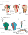
Organization of the wing imaginal disc. A 3rd instar wing disc viewed from the (A) columnar epithelium side (tan), (B) peripodial epithelium side (blue) and (C) in cross section. The A/P border (red) is contiguous on both surfaces. Hh (green) is expressed in all P compartment cells. Distributions of Hh, Dpp (ruby) and Wg (purple) in the wing primordium and peripodial epithelium, the disc-associated tracheal branch and myoblasts (orange) are shown in (A).
Figure 3
Figure 3
Models of Dpp and Bnl-FGF dispersion in wing discs. Columnar and peripodial cells are depicted in cross section, as is the disc-associated tracheal branch. Basal lamina encapsulates both disc and tracheal branch (gray). Dpp (red) is expressed at the developmental organizers/signaling centers of the columnar and peripodial layers (A-C); Bnl-FGF (black) is produced by columnar epithelial cells (D). Activation (yellow) is depicted in both disc and tracheal cells. (A) If Dpp is restricted to the epithelium surface, its activity is predicted to be restricted. (B) If Dpp moves freely after apical secretion, cross-lumenal signaling is predicted. (C) If Dpp moves freely after basal secretion, it is predicted to activate both disc and tracheal cells, since basally-secreted Bnl-FGF has been shown to signal through the basal lamina (D).
Figure 4
Figure 4
Four models of morphogen dispersion. Movement of morphogen (red) from source cell (middle, purple) to outlying cells by diffusion, serial transfer (transcytosis), lipoprotein particle transfer, and directly (via cytonemes).

References

    1. Martinez Arias A. Wnts as morphogens? The view from the wing of Drosophila. Nat Rev Mol Cell Biol. 2003;4:321–325. - PubMed
    1. Tabata T, Takei Y. Morphogens, their identification and regulation. Development. 2004;131:703–712. - PubMed
    1. Ashe HL, Briscoe J. The interpretation of morphogen gradients. Development. 2006;133:385–394. - PubMed
    1. Zhu AJ, Scott MP. Incredible journey: how do developmental signals travel through tissue? Genes Dev. 2004;18:2985–2997. - PubMed
    1. Eaton S. Release and trafficking of lipid-linked morphogens. Curr Opin Genet Dev. 2006;16:17–22. - PubMed

Publication types

Substances

LinkOut - more resources