Clinical Trial
. 2008 Mar;67(3):402-8.
doi: 10.1136/ard.2007.074229. Epub 2007 Jul 20.

Assessment of rituximab's immunomodulatory synovial effects (ARISE trial). 1: clinical and synovial biomarker results

Affiliations
Clinical Trial

Assessment of rituximab's immunomodulatory synovial effects (ARISE trial). 1: clinical and synovial biomarker results

A Kavanaugh et al. Ann Rheum Dis. 2008 Mar.

Abstract

Objective: Treatment with the anti-CD20 monoclonal antibody (mAb) rituximab is effective in rheumatoid arthritis (RA). Marked depletion of circulating B cells, seen in almost all patients, does not correlate with efficacy. The potential synovial immunomodulatory effects of rituximab have not been fully defined.

Methods: The ARISE trial is an open label, serial synovial biopsy (pre-treatment and 8 weeks) study of rituximab, given 1 g intravenously on days 0 and 14 without peri-infusional steroids, in active RA patients on concomitant methotrexate (MTX). Synovial tissue was analysed by immunohistochemistry with digital image analysis and gene expression by real-time PCR.

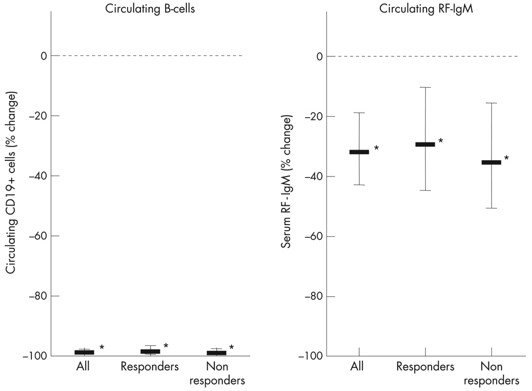
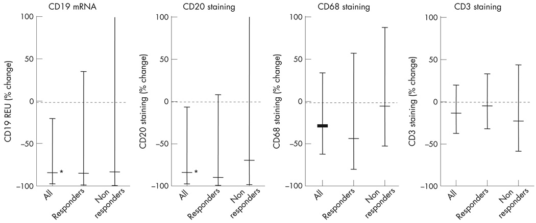
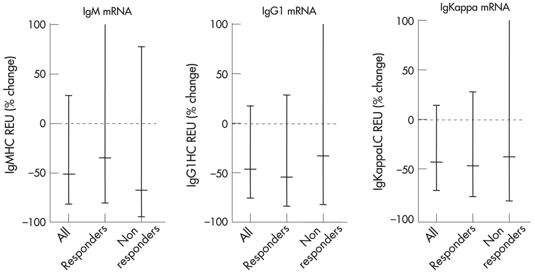
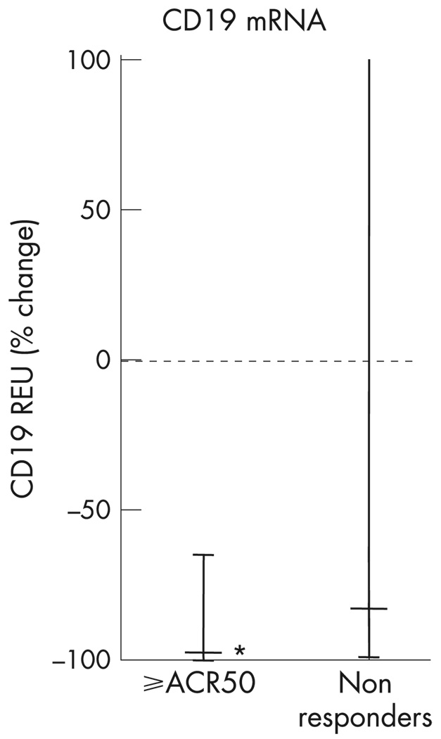
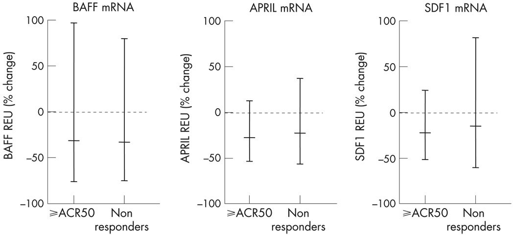
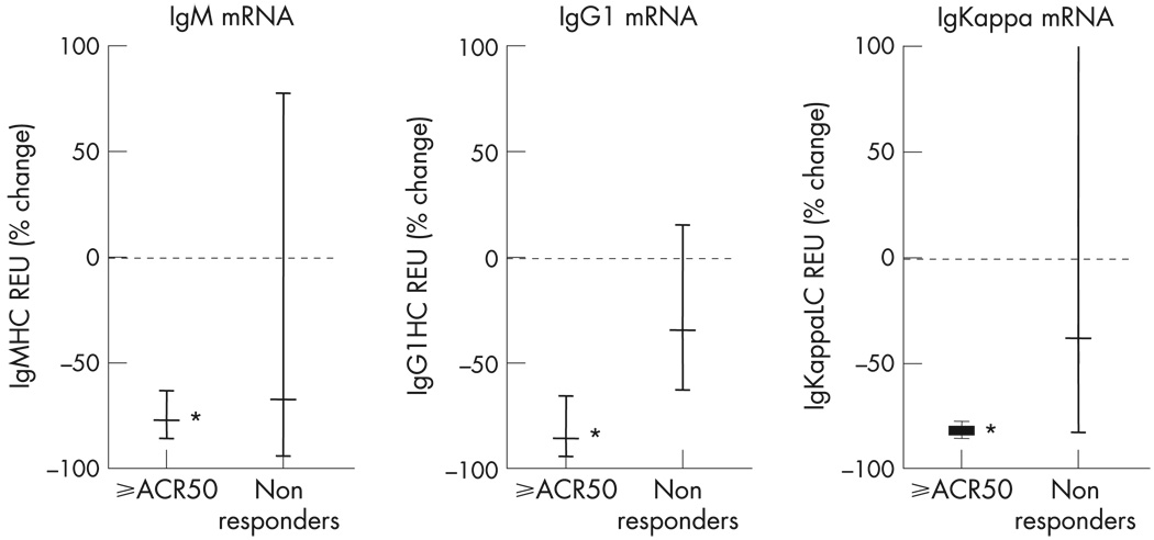
Results: The mean (SD) baseline DAS28 score was 6.5 (0.4), and mean MTX dose 17.3 mg/week. Of 13 patients, 11 had failed prior tumour necrosis factor (TNF) inhibitor therapy. With treatment, all patients experienced near complete depletion of circulating B cell numbers. During the 6 months after treatment, 7/13 patients achieved an American College of Rheumatology (ACR) 20% improvement (ACR20) response, 3/13 an ACR50 response and 2/13 an ACR70 response. There was a significant decrease in synovial B cells after treatment, but only a small trend towards greater reduction among clinical responders. Among the three patients with ACR50 responses there was a significant decrease in synovial immunoglobulin synthesis.

Conclusions: These data suggest that unlike those in circulation, synovial B cells are decreased but are not eliminated by rituximab therapy. Patients with higher levels of response may have more consistent depletion of synovial B cells, and may also have an alteration in synovial B cell function, as indicated by decreases in synovial immunoglobulin synthesis. Thus, effects on synovial B cells may be necessary but not sufficient for inducing clinical efficacy. Other effects, such as on primary lymph organ B cell antigen presentation or cytokine production, may be operative.


Figures

Figure 1
Figure 1
Mean Disease Activity Score (DAS)28 values over the initial 6 months for patients considered responders (n=7) or non-responders (n=6) to rituximab therapy are shown (error bars indicate standard deviations). This indicates the time course of clinical responses. Post-treatment biopsy was performed at week 8.
Figure 2
Figure 2
Changes in circulating B cells and rheumatoid factor (RF) after rituximab therapy. Data are shown as the percentage change in values from baseline to week 8. Data is shown for all patients, and separately for patients considered as responders and non-responders. Data represents geometric mean and 95% confidence intervals. *p<0.05.
Figure 3
Figure 3
Changes in various synovial cells after rituximab therapy. Data are shown as the percentage change in values from baseline to week 8. Data is shown for all patients, and separately for patients considered as responders and non-responders. Populations shown are B cells (CD19 and CD20); macrophages (CD68); T cells: CD3. Data represents geometric mean and 95% confidence intervals. *p<0.05.
Figure 4
Figure 4
Changes in synovial immunoglobulin synthesis after rituximab therapy. Data are shown as the percentage change in values from baseline to week 8. Data is shown for all patients, and separately for patients considered as responders and non-responders. Data represents geometric mean and 95% confidence intervals.
Figure 5
Figure 5
Changes in synovial inflammatory mediators after rituximab therapy. Data are shown as the percentage change in values from baseline to week 8. Data is shown for all patients, and separately for patients considered as responders and non-responders. Data represents geometric mean and 95% confidence intervals. *p<0.05.
Figure 6
Figure 6
Changes in synovial B cells after rituximab therapy. Data are shown as the percentage change in values from baseline to week 8. Data is shown for patients experiencing a 50% improvement in symptoms according to the American College of Rheumatology criteria (ACR50) or higher level of response during therapy (n=3) and for non-responders. Data represents geometric mean and 95% confidence intervals. *p<0.05.
Figure 7
Figure 7
Changes in synovial B cell survival factors after rituximab therapy. Data are shown as the percentage change in values from baseline to week 8. Data is shown for patients experiencing a 50% improvement in symptoms according to the American College of Rheumatology criteria (ACR50) or higher level of response during therapy (n=3) and for non-responders. Data represents geometric mean and 95% confidence intervals. *p<0.05.
Figure 8
Figure 8
Changes in synovial immunoglobulin synthesis after rituximab therapy. Data are shown as the percentage change in values from baseline to week 8. Data is shown for patients experiencing a 50% improvement in symptoms according to the American College of Rheumatology criteria (ACR50) or higher level of response during therapy (n=3) and for non-responders. Data represents geometric mean and 95% confidence intervals. *p<0.05.

References

    1. Edwards JCW, Cambridge G, Abrahams VM. Do self-perpetuating B lymphocytes drive human autoimmune disease? Immunology. 1999;97:188–196. - PMC - PubMed
    1. Kneitz C, Wilhelm M, Tony HP. Effective B cell depletion with rituximab in the treatment of autoimmune diseases. Immunobiology. 2002;206:519–527. - PubMed
    1. Silverman GJ, Weisman S. Rituximab therapy and autoimmune disorders: prospects for anti-B cell therapy. Arthritis Rheum. 2003;48:1484–1492. - PubMed
    1. Edwards JCW, Cambridge G. B-cell targeting in rheumatoid arthritis and other autoimmune diseases. Nat Rev Immunology. 2006;6:394–503. - PubMed
    1. Silverman GJ. Therapeutic B cell depletion and regeneration in rheumatoid arthritis: emerging patterns and paradigms. Arthritis Rheum. 2006;54:2356–2367. - PubMed

MeSH terms