. 2007 Sep;6(9):1528-37.
doi: 10.1128/EC.00185-07. Epub 2007 Jul 20.

Peroxisomal peripheral membrane protein YlInp1p is required for peroxisome inheritance and influences the dimorphic transition in the yeast Yarrowia lipolytica

Affiliations

Peroxisomal peripheral membrane protein YlInp1p is required for peroxisome inheritance and influences the dimorphic transition in the yeast Yarrowia lipolytica

Jinlan Chang et al. Eukaryot Cell. 2007 Sep.

Abstract

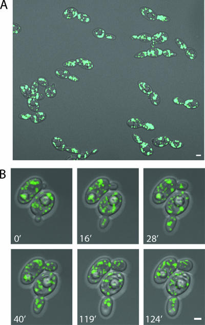
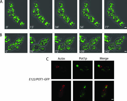
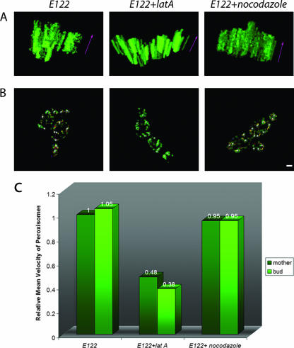
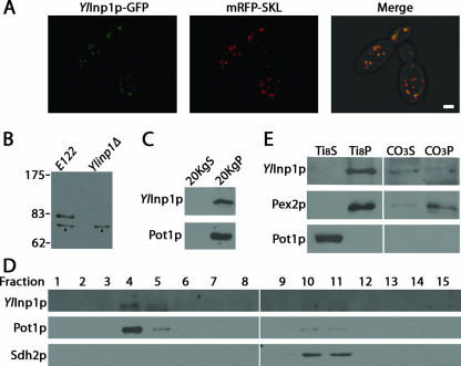
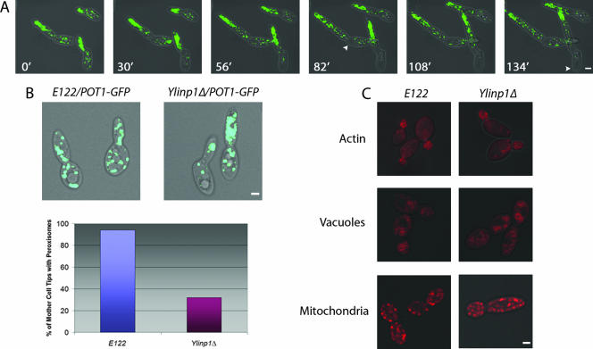
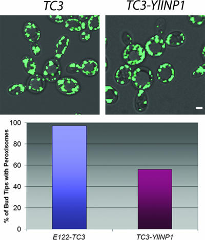
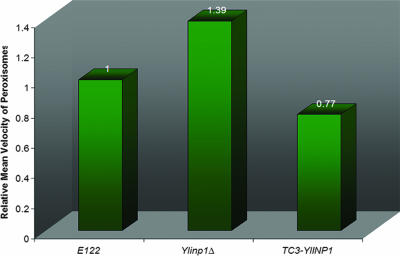
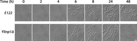
Eukaryotic cells have evolved molecular mechanisms to ensure the faithful inheritance of organelles by daughter cells in order to maintain the benefits afforded by the compartmentalization of biochemical functions. Little is known about the inheritance of peroxisomes, organelles of lipid metabolism. We have analyzed peroxisome dynamics and inheritance in the dimorphic yeast Yarrowia lipolytica. Most peroxisomes are anchored at the periphery of cells of Y. lipolytica. In vivo video microscopy showed that at cell division, approximately half of the anchored peroxisomes in the mother cell are dislodged individually from their static positions and transported to the bud. Peroxisome motility is dependent on the actin cytoskeleton. YlInp1p is a peripheral peroxisomal membrane protein that affects the partitioning of peroxisomes between mother cell and bud in Y. lipolytica. In cells lacking YlInp1p, most peroxisomes were transferred to the bud, with only a few remaining in the mother cell, while in cells overexpressing YlInp1p, peroxisomes were preferentially retained in the mother cell, resulting in buds nearly devoid of peroxisomes. Our results are consistent with a role for YlInp1p in anchoring peroxisomes in cells. YlInp1p has a role in the dimorphic transition in Y. lipolytica, as cells lacking the YlINP1 gene more readily convert from the yeast to the mycelial form in oleic acid-containing medium, the metabolism of which requires peroxisomal activity, than does the wild-type strain. This study reports the first analysis of organelle inheritance in a true dimorphic yeast and identifies the first protein required for peroxisome inheritance in Y. lipolytica.


Figures

FIG. 1.
FIG. 1.
Peroxisome dynamics in Y. lipolytica. (A) Wild-type cells expressing genomically encoded Pot1p-GFP to fluorescently label peroxisomes were grown in glucose-containing YPD medium and then transferred to oleic acid-containing YPBO medium and incubated for 16 h. Fluorescent images were captured by confocal microscopy. In this static picture, peroxisomes are seen to be evenly distributed between mother cell and bud. (B) The strain used in panel A was analyzed by 4D in vivo video microscopy. Representative frames from video S1 in the supplemental material show the specific movements of peroxisomes. One peroxisome already present in the small bud of the cell at bottom at the start of the video (0′) was observed later to return to the mother cell. At 16 min, one peroxisome in the cell at the top entered the bud. Additional peroxisomes entered this bud one by one between 16 min and 90 min. During this period, peroxisomes in the bud sometimes clustered at the center of the bud (28′) and later separated (40′), and some peroxisomes returned to the mother cell. Before cytokinesis, some peroxisomes from both the mother cell and bud relocated to the mother-bud neck region (119′) and then redistributed (124′). Bars, 2 μm.
FIG. 2.
FIG. 2.
Actin is involved in peroxisome dynamics. Treatment of cells with the actin-disrupting toxin latrunculin A (A) but not with the microtubule inhibitor nocodazole (B) abolished the dynamic movements of peroxisomes. Although cells grew more slowly following treatment with nocodazole, peroxisomes were still recruited to buds as normal. Representative frames from videos S2 and S3 in the supplemental material are presented in panels A and B, respectively. (C) Peroxisomes do not colocalize with actin patches. Wild-type cells synthesizing genomically encoded Pot1p-GFP were grown in YPD medium and then transferred to YPBO medium for 16 h. Actin was detected by staining with rhodamine-phalloidin and visualized by epifluorescence microscopy. Bar, 2 μm.
FIG. 3.
FIG. 3.
Quantification of peroxisome mobility. (A) One hundred projections corresponding to the last 20 min of videos S1, S2, and S3 in the supplemental material were analyzed with Imaris 4.1 (Bitplane), and 3D models were constructed. The z axis (purple arrows) represents time. A peroxisome that maintains its x-y position for the period of time considered and which is essentially immobile is represented by a fluorescent column parallel to the z axis. A mobile peroxisome is represented by fluorescent spots that have different x-y positions in time. (B) Tracking peroxisomes in untreated, latrunculin A-treated, and nocodazole-treated wild-type E122 cells. Randomly selected peroxisomes under each condition were tracked by analyzing the last 100 projections of videos S1, S2, and S3 with Imaris 4.1. The trajectories of individual peroxisomes are shown as different-colored lines. Bar, 2 μm. (C) Peroxisomes in latrunculin A-treated cells exhibit reduced mobility. The velocities of individual peroxisomes across individual time points were measured using Imaris 4.1, and an average velocity was obtained for each peroxisome. The average velocities of individual peroxisomes under a given condition were in turn averaged to obtain the mean velocity of peroxisomes under that condition. The mean velocity of peroxisomes under a given condition is expressed relative to the mean velocity of peroxisomes of the untreated wild-type E122/POT1-GFP strain, which was taken as 1.
FIG. 4.
FIG. 4.
Sequence alignment of S. cerevisiae Inp1p with the hypothetical YlInp1p encoded by the open reading frame YALI0F31229g of the Y. lipolytica genome. Amino acid sequences were aligned with the use of the ClustalW program (http://www.ebi.ac.uk/clustalw/; EMBL-EBI, Cambridgeshire, United Kingdom). Identical residues (black) and similar residues (gray) in the two proteins are shaded. Similarity rules: G = A = S; A = V; V = I = L = M; I = L = M = F = Y = W; K = R = H; D = E = Q = N; S = T = Q = N. Dashes represent gaps.
FIG. 5.
FIG. 5.
YlInp1p is a peripheral membrane protein of peroxisomes. (A) YlInp1p-GFP colocalizes with mRFP-SKL to punctate structures characteristic of peroxisomes by direct confocal microscopy. The right panel presents the merged image of the left and middle panels, with colocalization of YlInp1p-GFP and mRFP-SKL shown in yellow. Bar, 2 μm. (B) Immunoblot analysis of whole-cell lysates of the wild-type strain E122 and the deletion strain Ylinp1Δ probed with anti-YlInp1p antibodies. Strains were incubated in YPBO for 9 h. Arrowheads point to a nonspecific immunoreactive polypeptide present in the lysates of both E122 and Ylinp1Δ cells. (C) YlInp1p localizes to the 20KgP subcellular fraction enriched for peroxisomes. Immunoblot analysis of equivalent portions of the 20KgS and 20KgP subcellular fractions from wild-type E122 cells was performed with antibodies to YlInp1p and to the peroxisomal matrix enzyme thiolase (Pot1p). (D) YlInp1p cofractionates with peroxisomes. Organelles in the 20KgP fraction were separated by isopycnic centrifugation on a discontinuous sucrose gradient. Fractions were collected from the bottom of the gradient, and equal portions of each fraction were analyzed by immunoblotting. Fractions enriched for peroxisomes and mitochondria were identified by immunodetection of Pot1p and Sdh2p, respectively. (E) Purified peroxisomes were ruptured by treatment with Ti8 buffer and subjected to ultracentrifugation to obtain a supernatant fraction, Ti8S, enriched for matrix proteins and a pellet fraction, Ti8P, enriched for membrane proteins. The Ti8P fraction was treated further with alkali Na2CO3 and separated by ultracentrifugation into a supernatant fraction (CO3S) enriched for peripherally associated membrane proteins and a pellet fraction (CO3P) enriched for integral membrane proteins. Equivalent portions of each fraction were analyzed by immunoblotting. Immunodetection of Pot1p and Pex2p marked the fractionation profiles of a peroxisomal matrix and integral membrane protein, respectively.
FIG. 6.
FIG. 6.
Deletion of the YlINP1 gene affects specifically peroxisome inheritance. (A) Peroxisomes of the Ylinp1Δ strain were fluorescently labeled with genomically encoded Pot1p-GFP. Cells were grown for 16 h in YPD medium, transferred to YPBO medium for 16 h, and then visualized at room temperature with an LSM 510 META confocal microscope specially modified for 4D in vivo video microscopy (see Materials and Methods). Representative frames from video S4 in the supplemental material show the specific movements of peroxisomes in the Ylinp1Δ strain. At the start of the video (0′), cells already exhibit pseudohyphal characteristics, and peroxisomes are observed in mother cells and preferentially in buds. The cells at left show bidirectional growth. Continued video imaging showed that peroxisomes continue to move from mother cells to buds and localize to bud tips opposite mother cells (30′ to 133′). Mother cells are largely, but not completely, devoid of peroxisomes. Arrowheads point to tips of mother cells distal to the site of bud emergence that are devoid of peroxisomes. Bar, 2 μm. (B) The wild-type strain E122 and the deletion strain Ylinp1Δ expressing genomically integrated POT1-GFP were grown for 16 h in YPD medium and transferred to YPBO medium for 16 h. Fluorescence images were captured by confocal microscopy. Peroxisome inheritance was quantified as the percentage of mother cells retaining peroxisomes at their tips distal to the site of bud emergence. (C) Deletion of the YlINP1 gene does not affect actin structure or the inheritance of organelles other than peroxisomes. Wild-type E122 and Ylinp1Δ cells synthesizing Pot1p-GFP were grown in YPD medium. Actin was stained with rhodamine-phalloidin, vacuoles were stained with the fluorophore FM4-64, and mitochondria were stained with MitoTracker. Images were captured by confocal microscopy. Bar, 2 μm.
FIG. 7.
FIG. 7.
YlINP1 overexpression leads to peroxisome retention in mother cells. The strain E122/POT1-GFP was transformed with the empty plasmid pTC3 or with pTC3 containing the YlINP1 gene for overexpression of YlINP1. Cells were grown in YND medium and then transferred to and incubated in oleic acid-containing YNO medium for 16 h. Images were captured by confocal microscopy. Quantification of peroxisome retention by mother cells is reported as the percentage of bud tips containing peroxisomes. Bar, 2 μm.
FIG. 8.
FIG. 8.
Deletion or overexpression of the YlINP1 gene affects peroxisome mobility. The mean velocities of peroxisomes in wild-type E122/POT1-GFP, Ylinp1Δ/POT1-GFP, and YlINP1-overexpressing (TC3-YlINP1/POT1-GFP) cells were determined as described in the legend to Fig. 3.
FIG. 9.
FIG. 9.
Deletion of the YlINP1 gene affects the dimorphic transition. Wild-type E122 and Ylinp1Δ cells were grown in YPD medium for 16 h and then transferred to and incubated in YPBO medium. Samples were removed from YPBO at the times indicated and visualized by microscopy. Bar, 5 μm.

Similar articles

Cited by

References

    1. Davidson, R. C., J. R. Blankership, P. R. Kraus, M. de Jesus Berrios, C. M. Hull, C. D'Souza, P. Wang, and J. Heitman. 2002. A PCR-based strategy to generate integrative targeting alleles with large regions of homology. Microbiology 148:2607-2615. - PubMed
    1. de Duve, C., and P. Baudhuin. 1966. Peroxisomes (microbodies and related particles). Physiol. Rev. 46:323-357. - PubMed
    1. Fagarasanu, A., M. Fagarasanu, G. A. Eitzen, J. D. Aitchison, and R. A. Rachubinski. 2006. The peroxisomal membrane protein Inp2p is the peroxisome-specific receptor for the myosin V motor Myo2p of Saccharomyces cerevisiae. Dev. Cell 10:587-600. - PubMed
    1. Fagarasanu, M., A. Fagarasanu, and R. A. Rachubinski. 2006. Sharing the wealth: peroxisome inheritance in budding yeast. Biochim. Biophys. Acta 1763:1669-1677. - PubMed
    1. Fagarasanu, M., A. Fagarasanu, Y. Y. C. Tam, J. D. Aitchison, and R. A. Rachubinski. 2005. Inp1p is a peroxisomal membrane protein required for peroxisome inheritance in Saccharomyces cerevisiae. J. Cell Biol. 169:765-775. - PMC - PubMed

Publication types

LinkOut - more resources