. 2007 Oct;6(10):1853-64.
doi: 10.1128/EC.00088-07. Epub 2007 Jul 20.

Spitzenkorper localization and intracellular traffic of green fluorescent protein-labeled CHS-3 and CHS-6 chitin synthases in living hyphae of Neurospora crassa

Affiliations

Spitzenkorper localization and intracellular traffic of green fluorescent protein-labeled CHS-3 and CHS-6 chitin synthases in living hyphae of Neurospora crassa

Meritxell Riquelme et al. Eukaryot Cell. 2007 Oct.

Abstract

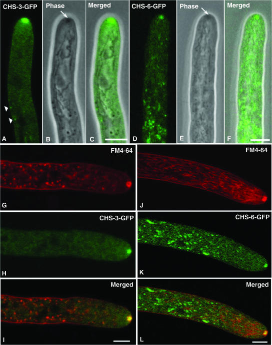
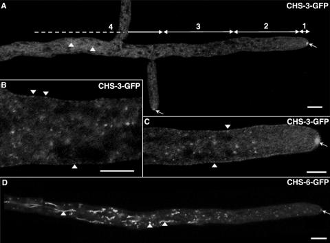
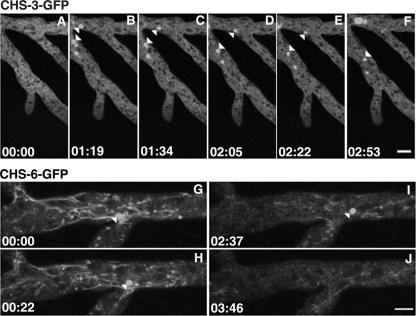
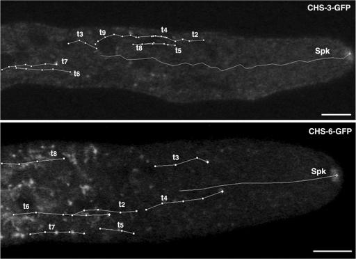
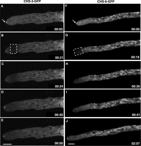
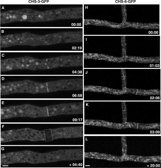
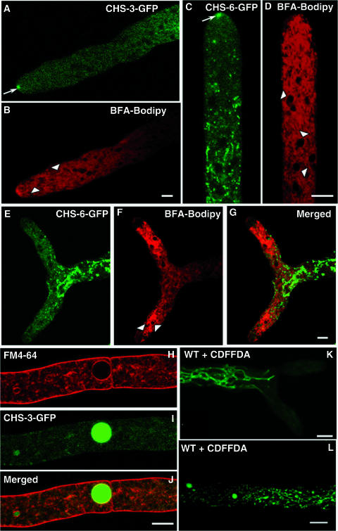
The subcellular location and traffic of two selected chitin synthases (CHS) from Neurospora crassa, CHS-3 and CHS-6, labeled with green fluorescent protein (GFP), were studied by high-resolution confocal laser scanning microscopy. While we found some differences in the overall distribution patterns and appearances of CHS-3-GFP and CHS-6-GFP, most features were similar and were observed consistently. At the hyphal apex, fluorescence congregated into a conspicuous single body corresponding to the location of the Spitzenkörper (Spk). In distal regions (beyond 40 microm from the apex), CHS-GFP revealed a network of large endomembranous compartments that was predominantly comprised of irregular tubular shapes, while some compartments were distinctly spherical. In the distal subapex (20 to 40 microm from the apex), fluorescence was observed in globular bodies that appeared to disintegrate into vesicles as they advanced forward until reaching the proximal subapex (5 to 20 microm from the apex). CHS-GFP was also conspicuously found delineating developing septa. Analysis of fluorescence recovery after photobleaching suggested that the fluorescence of the Spk originated from the advancing population of microvesicles (chitosomes) in the subapex. The inability of brefeldin A to interfere with the traffic of CHS-containing microvesicles and the lack of colocalization of CHS-GFP with the endoplasmic reticulum (ER)-Golgi body fluorescent dyes lend support to the idea that CHS proteins are delivered to the cell surface via an alternative route distinct from the classical ER-Golgi body secretory pathway.


Figures

FIG. 1.
FIG. 1.
Apical distribution of CHS-3-GFP and CHS-6-GFP in N. crassa (A to F) and comparison of CHS-GFP and FM4-64 (endocytic marker) localization (G to L). At the hyphal apex, fluorescence CSLM of GFP-labeled CHS-3 (A) and CHS-6 (D) shows a prominent apical body that coincides with the Spk, as revealed by phase-contrast microscopy (arrows in B, E) and confirmed in the overlaid images (C, F). Arrowheads in A point at fluorescent punctate structures at or near the plasma membrane; note the lack of correspondence with phase-dark structures in the merged image (C). Dual fluorescent labeling with FM4-64 (G, J) and CHS-3-GFP (H) or CHS-6-GFP (K) indicates no correspondence of labeled structures except for a concentric but only partial colocalization in the Spk. Overlaid images (I, L) show that the CHS-GFP occupies the center of a larger body stained by FM4-64. Scale bars, 5 μm.
FIG. 2.
FIG. 2.
Distribution of GFP-labeled CHS-3 and CHS-6 along hyphae. Reconstruction of a Neurospora hypha, showing overall distribution of CHS-3-GFP (A). Hyphae were divided into four regions measured from the tip: 1, apex (2 to 5 μm); 2, proximal subapex (5 to 20 μm); 3, distal subapex (20 to 40 μm); and 4, distal region (>40 μm). In the distal (B) and proximal subapex (C) CHS-3-GFP shows a punctate distribution; some fluorescent dots appear to be at the plasma membrane (arrowheads in panel B, C). Reconstruction of a hypha showing overall distribution of CHS-6-GFP (D). In distal regions, both CHS-3-GFP and CHS-6-GFP are found in a highly stained network of spherical and tubular compartments (arrowheads in panel A, D). Arrows in panels A, C, and D point to the Spk. Scale bars: 10 μm (A, D); 5 μm (B, C).
FIG. 3.
FIG. 3.
Progressive disintegration of GFP-labeled vacuoles and dispersal of fluorescence in strain NMR3 (A to F) and strain NMR6 (G to J). Arrowheads point at spherical bodies whose size gradually diminishes until they are no longer visible. Similarly, the CHS-6-GFP-labeled tubular compartments in NMR6 undergo progressive disintegration (G to J). Time, min:s. Scale bars, 5 μm.
FIG. 4.
FIG. 4.
Kinetic analysis of fluorescent structures carrying CHS-3-GFP and CHS-6-GFP. The trajectories (t) of individual particles and the Spk were followed. Fluorescent vesicles were traced moving predominantly forward until reaching the proximal subapex (15 to 20 μm from the apex) where they were no longer visible. The average speed (in μm/s) of the Spk and the different vesicles was, for strain NMR3 Spk, 0.11 ± 0.02; t2, 0.11 ± 0.06; t3, 0.13 ± 0.04; t4, 0.12 ± 0.09; t5, 0.11 ± 0.04; t6, 0.18 ± 0.05; t7, 0.12 ± 0.04; t8, 0.13 ± 0.04; and t9, 0.09 ± 0.03; and for strain NMR6 Spk, 0.24 ± 0.02; t2, 0.18 ± 0.05; t3, 0.19 ± 0.06; t4, 0.21 ± 0.02; t5, 0.18 ± 0.07; t6, 0.26 ± 0.02; t7, 0.19 ± 0.06; and t8, 0.22 ± 0.05. Time, min:s. Scale bars, 5 μm. Particle movement can be better appreciated in movies S1 and S2 in the supplemental material.
FIG. 5.
FIG. 5.
FRAP analysis of the proximal subapex of a CHS-3-GFP hypha (A to E) and the apex of a CHS-6-GFP hypha (F to J). Prebleached hyphae. Arrows point at the Spk (A, F). Photobleaching was applied to the selected areas indicated by the dotted rectangles (B, G). Recovery of Spk fluorescence occurs after either apical (H to J) or subapical (C to E) bleaching. Note a temporary departure of the Spk from its centric position in the apical dome (D). Time, min:s. Scale bars, 5 μm.
FIG. 6.
FIG. 6.
Localization of CHS-GFP during cross-wall synthesis. CSLM time series show the progressive and localized accumulation of both CHS-3-GFP (A to E) and CHS-6-GFP (H to K) during septum formation. In a FRAP analysis, the formed septum disappeared after photobleaching (F) and no fluorescence reappeared in the septum even after 4 min following FRAP (G). Septum completion in strain NMR6 lasts ∼3 to 6 min (H to K); afterwards, the CHS-6-GFP signal dissipates over time; 20 minutes after the septum had been completed, septum fluorescence had vanished (L). Time, min:s. Scale bars, 5 μm.
FIG. 7.
FIG. 7.
Distinct localization of CHS-GFP and conventional secretory compartments. (A to G) Comparative distribution of fluorescence from CHS-3-GFP (A) and CHS-6-GFP (C, E) with Golgi body structures revealed after staining with 5 μM BFA-bodipy 558/568 conjugate. After a short period (∼5 min), the dye stained apical and subapical Golgi body equivalents (arrowheads in B). Note the preservation of CHS-GFP in the Spk (arrow in panel A). Over 5 to 20 min, the dye stained the ER (arrowheads in D). During that period, GFP fluorescence was still very prominent at the Spk of strain NMR6 (arrow in panel C). After a longer exposure (20 to 25 min), the hyphae stopped growing (E to G), the Spk disappeared, and both fluorescence and dye moved away from the apical region and strongly stained the ER (arrowheads in panel F), but the separation between fluorescent CHS-6-GFP and the bodipy-stained structures, particularly the ER around the nuclei, remained clearly evident (G). Scale bars, 5 μm. (H to L) Relationship between CHS-3-GFP and the vacuolar system. Localization of CHS-3-GFP fluorescence in the vacuoles of NMR3 in hyphae stained with FM4-64 (H to J); note that CHS-3-GFP is found in the lumen of the vacuole (I), whereas FM4-64 stains the vacuolar membrane (H, J). Scale bars, 5 μm. For comparison, the vacuolar system in wild-type hyphae of N. crassa is shown after staining with 10 μM CDFFDA (K, L). Scale bars, 10 μm.

Similar articles

Cited by

References

    1. Amnuaykanjanasin, A., and L. Epstein. 2006. A class Vb chitin synthase in Colletotrichum graminicola is localized in the growing tips of multiple cell types, in nascent septa, and during septum conversion to an end wall after hyphal breakage. Protoplasma 227:155-164. - PubMed
    1. Bartnicki-Garcia, S. 1968. Cell wall chemistry, morphogenesis and taxonomy of fungi. Annu. Rev. Microbiol. 22:87-107. - PubMed
    1. Bartnicki-Garcia, S. 2006. Chitosomes: past, present and future. FEMS Yeast Res. 6:957-965. - PubMed
    1. Bartnicki-Garcia, S. 2002. Hyphal tip growth: outstanding questions, p. 29-58. In H. D. Osiewacz (ed.), Molecular biology of fungal development. Marcel Dekker, Inc., New York, NY.
    1. Bartnicki-Garcia, S. 1990. Role of vesicles in apical growth and a new mathematical model of hyphal morphogenesis, p. 211-232. In I. B. Heath (ed.), Tip growth in plant and fungal cells. Academic Press, San Diego, CA.

Publication types

MeSH terms

LinkOut - more resources