. 2007 Jul;19(7):2264-77.
doi: 10.1105/tpc.106.045732. Epub 2007 Jul 20.

Functional analysis of the epidermal-specific MYB genes CAPRICE and WEREWOLF in Arabidopsis

Affiliations

Functional analysis of the epidermal-specific MYB genes CAPRICE and WEREWOLF in Arabidopsis

Rumi Tominaga et al. Plant Cell. 2007 Jul.

Abstract

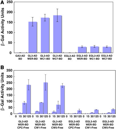
Epidermis cell differentiation in Arabidopsis thaliana is a model system for understanding the developmental end state of plant cells. Two types of MYB transcription factors, R2R3-MYB and R3-MYB, are involved in cell fate determination. To examine the molecular basis of this process, we analyzed the functional relationship of the R2R3-type MYB gene WEREWOLF (WER) and the R3-type MYB gene CAPRICE (CPC). Chimeric constructs made from the R3 MYB regions of WER and CPC used in reciprocal complementation experiments showed that the CPC R3 region cannot functionally substitute for the WER R3 region in the differentiation of hairless cells. However, WER R3 can substantially substitute for CPC R3. There are no differences in yeast interaction assays of WER or WER chimera proteins with GLABRA3 (GL3) or ENHANCER OF GLABRA3 (EGL3). CPC and CPC chimera proteins also have similar activity in preventing GL3 WER and EGL3 WER interactions. Furthermore, we showed by gel mobility shift assays that WER chimera proteins do not bind to the GL2 promoter region. However, a CPC chimera protein, which harbors the WER R3 motif, still binds to the GL2 promoter region.


Figures

Figure 1.
Figure 1.
WER (R2R3 MYB) and CPC (R3 MYB) Genes in Arabidopsis. (A) Sequence alignment of the WER and CPC proteins. Shaded letters indicate identical residues. The MYB DNA binding domains present in each of these proteins are indicated. Although WER has two MYB domains (R2 and R3), CPC has only R3. The positions of the three helices (h) forming R3 MYB are shown with green lines. (B) Helical diagrams of helix 1, helix 2, and helix 3 in WER R3 and CPC R3 with nonpolar residues in yellow, polar uncharged residues in green, acidic residues in red, and basic residues in blue.
Figure 2.
Figure 2.
Phylogenetic Tree Displaying the Relationship among R3 Myb Regions. (A) A neighbor-joining phylogenetic tree of the amino acid sequences of R3 Myb regions (CPC, TRY, ETC1, ETC2, At4g01060, WER, MYB23, GL1, MYB36, MYB37, MYB38, MYB68, MYB84, MYB87, PAP1, and PAP2). Distances are shown as the p-distance multiplied by 103. Branches with bootstraps of 90% or greater are in bold. Branches with bootstraps between 70 and 90% are marked with a circle. Branches with bootstraps below 70% are unmarked. (B) Amino acid sequences of Myb R3 motifs of CPC, TRY, ETC1, ETC2, At4g01060 WER, GL1, MYB23, MYB36, MYB37, MYB38, MYB68, MYB84, MYB87, PAP1, and PAP2. Boxes outlined in red indicate identical amino acids.
Figure 3.
Figure 3.
Complementation of the wer Mutant Phenotype by WER:WER-CPC Chimera Constructs. (A) Schematic representation of chimera WER R3 (yellow) and CPC R3 (red) constructs. Complementation results are on the right. Numbers indicate the amino acids removed from WER regions as indicated in (B). Only WER:WER and WER:WC2 could complement the wer mutant phenotype. (B) Alignment of the MYB R3 regions of WER and CPC. Shaded letters indicate identical residues. The positions of the three helices forming R3 MYB are indicated with green lines. (C) Phenotypes of Col-0, wer, and wer transformants. Transformants with WER:WER and WER:WC2 had a decreased number of root hairs compared with wer. Bar = 100 μm.
Figure 4.
Figure 4.
Regulation of GL2:GUS in the wer Mutant Background. Expression of the GL2:GUS reporter in the developing root epidermis of 5-d-old seedlings in Col-0, wer, and wer transformants. GL2 promoter activity is reduced in the epidermis of the wer line. Transformants WER:WER and WER:WC2 had increased GL2 promoter activity compared with wer. Bar = 100 μm.
Figure 5.
Figure 5.
Complementation of the cpc Mutant by CPC:CPC-WER Chimera Constructs. (A) Schematic representation of chimera CPC R3 (red) and WER R3 (yellow) constructs. Complementation results are on the right. Each of the constructs complemented the cpc mutant phenotype. Numbers indicate replaced WER regions as shown in (B). (B) Alignment of the MYB R3 regions of WER and CPC. Shaded letters indicate identical residues. The positions of the three helices forming R3 MYB are shown with green lines. (C) Phenotypes of Col-0, cpc, and cpc transformants. All transgenic plant lines had an increased number of root hairs compared with cpc. Bar = 100 μm.
Figure 6.
Figure 6.
Regulation of GL2:GUS in the cpc Mutant Background. Expression of the GL2:GUS reporter in the developing root epidermis of 5-d-old seedlings in Col-0, cpc, and cpc transformants. GL2 promoter activity is increased in the epidermis of the cpc line. All transformant lines had reduced GL2 promoter activity compared with cpc with overnight incubation in X-Gluc solution. Bar = 100 μm.
Figure 7.
Figure 7.
Protein Interactions with Native Myb versus Chimeric Myb Proteins. (A) Comparison of protein interactions using a yeast two-hybrid assay between WER, WC1, and WC7 with GL3 and EGL3. The WER, WC1, and WC7 proteins were compared as GAL4 binding domain (BD) fusions, whereas GL3 and EGL3 were expressed as GAL4 activation domain (AD) fusions. (B) CPC, CW1, and CW5 competition for the WER binding sites of GL3 and EGL3. Using the pBridge vector (Clontech), a third protein (CPC, CW1, or CW5) under the control of a Met-repressible promoter was expressed in a yeast interaction assay at varying Met concentrations (0, 15, 30, and 125 μM). CPC, CW1 or CW5 were expressed as a free protein (no AD or BD domains). Samples were normalized by OD550. The strength of the interaction was determined by β-gal activity.
Figure 8.
Figure 8.
The DNA Binding Properties of WER and CPC Chimera Proteins. (A) WER, WC1, or WC7 protein was added with or without a 200-fold excess of competitor. (B) CPC or CW5 protein was added with or without a 200-fold excess of competitor. Arrows indicate shifted bands, and arrowheads indicate free probe. Digoxigenin-labeled DNA of GL2MBS1 was used as the probe.
Figure 9.
Figure 9.
Regulatory Cascade Models for WER and CPC Chimeras. (A) WER and CPC proteins competitively bind to the GL3/EGL3-TTG1 complex. (B) The WER-GL3/EGL3-TTG1 complex can bind the GL2 promoter to promote GL2 expression, which leads to the hairless cell fate, whereas the WER chimera-GL3/EGL3-TTG1 complex cannot bind to the GL2 promoter, thus preventing GL2 expression. (C) Both CPC-GL3/EGL3-TTG1 and CPC chimera-GL3/EGL3-TTG1 complexes prevent expression of GL2. The absence of GL2 results in root hair formation.
Figure 10.
Figure 10.
Evolutionary Models of CPC and WER. (A) WER binds the GL2 promoter to promote GL2 expression. (B) Proto-CPC protein derived from WER (yellow) truncation can bind the promoter but prevents expression of GL2. (C) CPC cannot bind to the GL2 promoter, strongly preventing GL2 expression. (D) Proto-CPC protein derived from WER amino acid substitution cannot bind to the GL2 promoter.

References

    1. Andersson, K.B., Berge, T., Matre, V., and Gabrielsen, O.S. (1999). Sequence selectivity of c-Myb in vivo. Resolution of a DNA target specificity paradox. J. Biol. Chem. 274 21986–21994. - PubMed
    1. Ausubel, F., Brent, R., Kingston, R.E., Moore, D.D., Seidman, J.G., Smith, J.A., and Struhl, K. (1995). Current Protocols in Molecular Biology. (New York: John Wiley & Sons).
    1. Bechtold, N., Ellis, J., and Pelletier, G. (1993). In planta Agrobacterium-mediated gene transfer by infiltration of adult Arabidopsis thaliana plants. C. R. Acad. Sci. Paris 316 1194–1199.
    1. Bernhardt, C., Lee, M.M., Gonzalez, A., Zhang, F., Lloyd, A., and Schiefelbein, J. (2003). The bHLH genes GLABRA3 (GL3) and ENHANCER OF GLABRA3 (EGL3) specify epidermal cell fate in the Arabidopsis root. Development 130 6431–6439. - PubMed
    1. Bernhardt, C., Zhao, M., Gonzalez, A., Lloyd, A., and Schiefelbein, J. (2005). The bHLH genes GL3 and EGL3 participate in an intercellular regulatory circuit that controls cell patterning in the Arabidopsis root epidermis. Development 132 291–298. - PubMed

MeSH terms