

Is the prefrontal bone in Alpine newt (Triturus alpestris Laurenti, 1768) of dual origin?
- PMID: 17645458
- PMCID: PMC2375820
- DOI: 10.1111/j.1469-7580.2007.00768.x
Is the prefrontal bone in Alpine newt (Triturus alpestris Laurenti, 1768) of dual origin?
Abstract
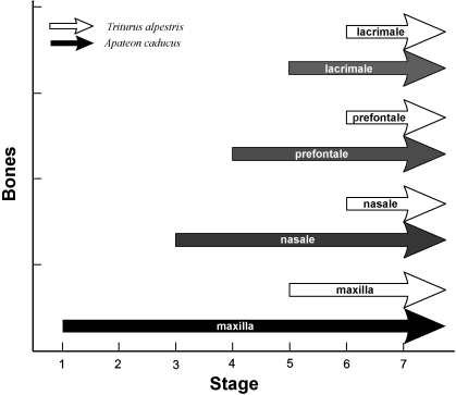
According to current knowledge, only the prefrontal bone (os prefrontale) of the circumorbital series is preserved in the family Salamandridae. However, the exact origin and number of ossification centres creating this bone is unknown. Detailed examination of the prefrontal bone during ontogeny of juvenile and adult specimens of the Alpine Newt (Triturus alpestris) indicates its dual origin (prefrontal and lacrimal). We found that the prefrontal bone originates from four ossification centres, i.e. three prefrontal centres and one posterior lacrimal centre. The anterior lacrimal centre participates in the maxillar ossification. The development of these ossification centres occurs very late in ontogeny (at stage 54), and starts after differentiation of the nasal capsules. The total fusion of the lacrimal ossification centre with the prefrontal bone of T. alpestris is distinct from the fully differentiated lacrimal bone attached to the prefrontal bone of the fossil family Branchiosauridae (Temnospondyly). We propose that heterochrony, observed in the recent species, is a delayed development followed by accelerated ossification that resulted in the fusion of the anterior lacrimal centre with the maxilla and the posterior lacrimal centre with the prefrontal bone.
Figures


Similar articles
-
Bony skull of the smooth newt Triturus vulgaris (Amphibia, Urodela: Salamandridae) and the role of thyroid hormones in its ossification.Dokl Biol Sci. 2003 Jan-Feb;388:73-5. doi: 10.1023/a:1022416530987. Dokl Biol Sci. 2003. PMID: 12705137 No abstract available.
-
Skull development in the Iberian newt, Pleurodeles waltl (Salamandridae: Caudata: Amphibia): timing, sequence, variations, and thyroid hormone mediation of bone appearance.J Anat. 2020 Sep;237(3):543-555. doi: 10.1111/joa.13210. Epub 2020 May 15. J Anat. 2020. PMID: 32412118 Free PMC article.
-
The development of the larval pigment patterns in Triturus alpestris and Ambystoma mexicanum.Adv Anat Embryol Cell Biol. 1990;118:1-99. Adv Anat Embryol Cell Biol. 1990. PMID: 2368640
-
Paedogenesis in european newts (Triturus: salamandridae): cranial morphology during ontogeny.J Morphol. 2000 Feb;243(2):127-39. doi: 10.1002/(SICI)1097-4687(200002)243:2<127::AID-JMOR2>3.0.CO;2-0. J Morphol. 2000. PMID: 10658197
-
Integration of the sensory and skeletal systems: A classical perspective on neuromast-bone interactions.Dev Biol. 2025 Aug;524:48-54. doi: 10.1016/j.ydbio.2025.04.012. Epub 2025 Apr 30. Dev Biol. 2025. PMID: 40315947 Review.
Cited by
-
Stem caecilian from the Triassic of Colorado sheds light on the origins of Lissamphibia.Proc Natl Acad Sci U S A. 2017 Jul 3;114(27):E5389-E5395. doi: 10.1073/pnas.1706752114. Epub 2017 Jun 19. Proc Natl Acad Sci U S A. 2017. PMID: 28630337 Free PMC article.
-
A New Basal Salamandroid (Amphibia, Urodela) from the Late Jurassic of Qinglong, Hebei Province, China.PLoS One. 2016 May 4;11(5):e0153834. doi: 10.1371/journal.pone.0153834. eCollection 2016. PLoS One. 2016. PMID: 27144770 Free PMC article.
References
-
- Alberch P, Gould SJ, Oster GF, Wake DB. Size and shape in ontogeny and phylogeny. Paleobiology. 1979;5:296–317.
-
- Balon EK, Fleger-Balon C. Microscopic techniques for studies of early ontogeny in fishes: problems and methods of composite descriptions. In: Balon EK, editor. Early Life Histories of Fishes. Dordrecht: Dr W. Junk Publishers; 1985. pp. 33–55.
-
- Boy J. Die Branchiosaurier (Amphibia) des saarpfälzischen Rotliegenden (Perm, SW – Deutschland) Abh Hessisch Landesamt Bodenforsch. 1972;65:1–137.
-
- Boy JA, Sues H-D. Branchiosuars: Larvae, metamorphosis and heterochrony in temnospondyls and seymouriamorphs. In: Healtwole H, Carroll RL, editors. Amphinbian Biology, – Palaeontology. Vol. 4. Chipping Norton: Surrey Beatty & Sons; 2000. pp. 1150–1197.
-
- Corsin J. The development of the osteocranium of Pleurodeles waltlii Michahelles. J Morph. 1966;119:209–216. - PubMed
MeSH terms
LinkOut - more resources
Full Text Sources
