. 2007 Sep;8(9):1280-96.
doi: 10.1111/j.1600-0854.2007.00610.x. Epub 2007 Jul 23.

Targeting of Sna3p to the endosomal pathway depends on its interaction with Rsp5p and multivesicular body sorting on its ubiquitylation

Affiliations

Targeting of Sna3p to the endosomal pathway depends on its interaction with Rsp5p and multivesicular body sorting on its ubiquitylation

Marta Stawiecka-Mirota et al. Traffic. 2007 Sep.

Abstract

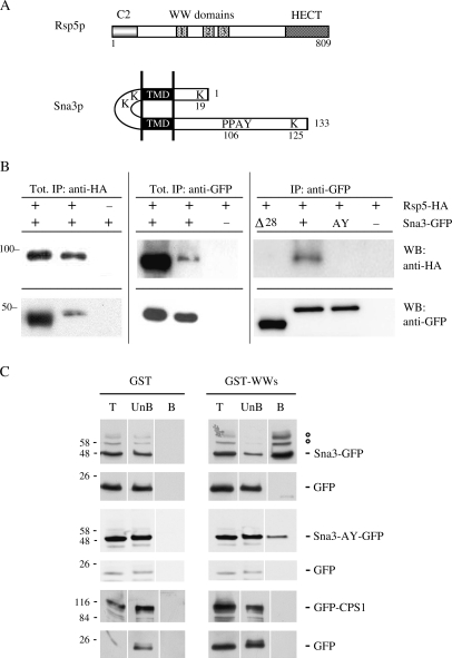
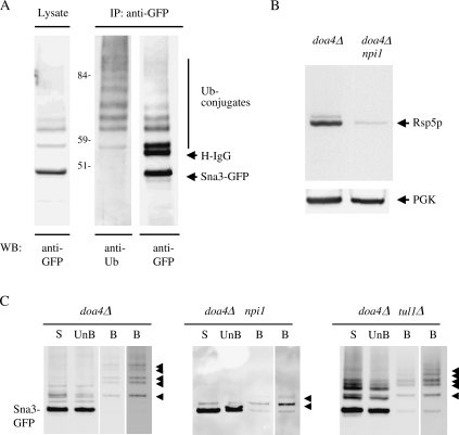
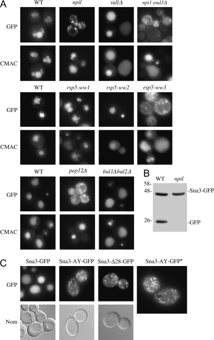
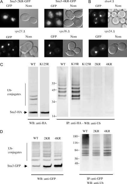
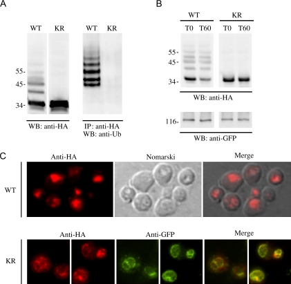
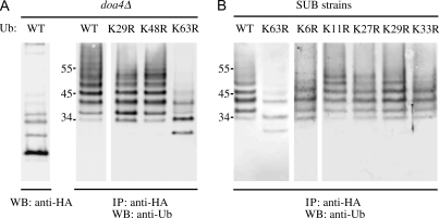
Rsp5p is an ubiquitin (Ub)-protein ligase of the Nedd4 family that carries WW domains involved in interaction with PPXY-containing proteins. It plays a key role at several stages of intracellular trafficking, such as Ub-mediated internalization of endocytic cargoes and Ub-mediated sorting of membrane proteins to internal vesicles of multivesicular bodies (MVBs), a process that is crucial for their subsequent targeting to the vacuolar lumen. Sna3p is a membrane protein previously described as an Ub-independent MVB cargo, but proteomic studies have since shown it to be an ubiquitylated protein. Sna3p carries a PPXY motif. We observed that this motif mediates its interaction with Rsp5p WW domains. Mutation of either the Sna3p PPXY motif or the Rsp5p WW3 domain or reduction in the amounts of Rsp5 results in the mistargeting of Sna3p to multiple mobile vesicles and prevents its sorting to the endosomal pathway. This sorting defect appears to occur prior to the defect displayed in rsp5 mutants by other MVB cargoes, which are correctly sorted to the endosomal pathway but missorted to the vacuolar membrane instead of the vacuolar lumen. Sna3p is polyubiquitylated on one target lysine, and a mutant Sna3p lacking its target lysine displays defective MVB sorting. Sna3p undergoes Rsp5-dependent polyubiquitylation, with K63-linked Ub chains.


Figures

Figure 1
Figure 1
Binding of the PY motif of Sna3p to the WW domains of Rsp5p.A) Schematic representation of Rsp5p and Sna3p. B) Immunoprecipitation was performed with 170 μg of total extracts from cells producing HA-tagged Rsp5p under the control of its own promoter and transformed by a plasmid encoding GFP-tagged Sna3p wild type or mutants under the control of TPI1 promoter. Tagged proteins were immunoprecipitated with anti-HA or anti-GFP antibodies. Cells producing untagged Rsp5p or Sna3p were used as controls. Immunoprecipitated proteins were detected by immunoblotting with anti-HA and anti-GFP antibodies. Tot: 6% of the solubilized proteins were loaded on the gel. IP: 50% of the immunoprecipitated material was loaded on the gel. C) Glutathione S-transferase and GST-WW2/WW3 Rsp5p domain recombinant proteins bound to glutathione–Sepharose beads were incubated with extracts from cells producing Sna3-GFP, Sna3-AY-GFP or GFP-Cps1p. Total extract (T), unbound (UnB) and bound (B) fractions were analyzed by Western blotting with anti-GFP. °: These bands were demonstrated to be ubiquitylated Sna3p species (see later). IP: immunoprecipitation, WB: western-blot.
Figure 3
Figure 3
Sna3p is ubiquitylated by the E3 ligase Rsp5p.A) Green fluorescent protein-tagged version of Sna3p was detected either by immunoblotting of a cell lysate from wild-type (WT) cells with anti-GFP or by immunoblotting with anti-Ub or anti-GFP after immunoprecipitation with anti-GFP. Molecular weight markers sizes are indicated. B) Rsp5p and PGK, a loading control protein, were detected by immunoblotting of a cell lysate from doa4Δ and doa4Δnpi1 cells with anti-mNedd4 and anti-PGK antibodies, respectively. C) doa4Δ, doa4Δnpi1 and doa4Δtul1Δ cells co-producing Sna3-GFP and CUP1 promoter-driven 6His-Ub were grown to mid-exponential phase. CuSO4 (100 μm) was added, and the cells were incubated for an additional 3 h to induce the CUP1 promoter. Extracts of 6 × 10⁸ to 7 × 10⁸ cells were fractionated as described in Materials and Methods. All experiments were conducted in identical conditions of growth and cell fractionation in at least three independent experiments. Solubilized membranes were passed through Ni-NTA columns. The unbound fractions were collected and the 6His-tagged ubiquitylated proteins were then eluted using a buffer containing 200 mm imidazole. Aliquots of each fraction were resolved by electrophoresis and analyzed by Western immunoblotting with anti-GFP antibodies. Solubilized membrane fractions (S), unbound fraction (UnB) and purified 6His-tagged proteins (B) are shown. The lane on the right is overexposed. Arrowheads indicate Sna3-GFP conjugated to 6His-tagged Ub moieties. IP: immunoprecipitation, WB: western-blot.
Figure 2
Figure 2
Targeting of Sna3-GFP to the vacuolar lumen is abolished in rsp5 mutant cells and if the Sna3p PY motif is altered.Microscopic images of Sna3-GFP in living cells. Cells transformed by a Sna3-GFP plasmid under the control of the TPI1 promoter were grown to mid-exponential growth phase and the fluorescence was examined under the microscope. A) Green fluorescent protein and CMAC staining in 27061b parental strain and its derivatives, npi1, tul1Δ and npi1 end3Δ mutant cells; in the BG1 parental strain and derivative cells deleted for RSP5 and producing a centromeric plasmid version of Rsp5p wild type (WT) or mutated in the WW1, WW2 or WW3 domain; and in BY parental strain, pep12Δ and bul1Δbul2Δ mutant cells. The identity of the vacuole was confirmed by staining with the dye CMAC as described in Materials and Methods. B) Western blots from total protein extracts from WT and npi1 mutant cells producing Sna3-GFP were probed with anti-GFP antibodies. The sizes of molecular weight markers are indicated. C) Cells transformed by a plasmid encoding GFP-tagged WT or Sna3p variants were grown to mid-exponential phase and cells were examined for fluorescence and with Nomarski (Nom) optics. *, the image of Sna3-AY-GFP is magnified.
Figure 4
Figure 4
Sna3p is polyubiquitylated on one target lysine, K125.A) Cells expressing Sna3-K19,125R-GFP and Sna3-4KR-GFP were grown to mid-exponential growth phase, and cells were examined for fluorescence and with Nomarski (Nom) optics. B) doa4Δ, vps23Δ, vps36Δ and vps24Δ cells transformed by a Sna3-GFP encoding plasmid were grown to mid-exponential growth phase, and cells were examined for fluorescence and with Nomarski optics. C) Left panel: HA-tagged version of Sna3p and Sna3-K125R were detected by immunoblotting of corresponding cell lysates from wild-type (WT) cells with anti-HA antibodies. Right panel: Modified forms of Sna3-HA and its KR mutant derivatives were detected by immunoblotting with anti-Ub antibodies after immunoprecipitation with anti-HA antibodies. D) Modified forms of Sna3-GFP and its KR mutant derivatives were detected by immunoblotting with anti-GFP antibodies or by immunoblotting with anti-Ub antibodies after immunoprecipitation with anti-GFP. The sizes of molecular weight markers are indicated. IP: immunoprecipitation, WB: western-blot.
Figure 5
Figure 5
Sna3p undergoes Ub-dependent MVB sorting.sna3Δand VPH1-GFP cells expressing Sna3p-6HA or Sna3p-4KR-6HA were grown to mid-exponential phase. A) Sna3p-6HA or Sna3p-4KR-6HA produced in sna3Δ cells were detected by immunoblotting of cell lysates with anti-HA antibodies (left panel) and immunoblotting with anti-Ub antibodies after immunoprecipitation with anti-HA antibodies (right panel). B) The production of Sna3p-6HA or Sna3p-4KR-6HA in VPH1-GFP cells was stopped by adding cycloheximide (100 μg/mL). Sna3p-6HA or Sna3p-4KR-6HA was detected by immunoblotting of cell lysates with anti-HA antibodies at the times indicated after the addition of cycloheximide, while Vph1-GFP was detected with anti-GFP antibodies. C) Phenylmethylsulfonyl fluoride was added for 1 h toVPH1-GFP cells producing Sna3p-6HA or Sna3p-4KR-6HA before the cells were fixed and processed for immunofluorescence with monoclonal anti-HA and polyclonal anti-GFP antibodies followed by Texas Red-labeled donkey anti-mouse IgG and FITC-labeled donkey anti-rabbit IgG as described in Materials and Methods. Cells were examined for fluorescence (the HA signal was detected using the rhodamine filter set and the GFP signal using the FITC filter set) and vacuole by Nomarski optics. IP: immunoprecipitation, WB: western-blot.
Figure 6
Figure 6
Sna3p is modified by Lys63-linked Ub chains.A) doa4Δ cells co-transformed with Sna3p-HA encoding plasmid and plasmids encoding wild-type (wt) Ub, UbK29R, UbK48R or UbK63R were grown to mid-exponential phase and induced for Ub synthesis for 1 h as described in Figure 3C. Modified forms of Sna3-HA and its KR mutant derivatives were detected by immunoblotting with anti-Ub antibodies after immunoprecipitation with anti-HA. The positions of size markers are indicated. B) Sna3-HA ubiquitylation in cells whose sole source of Ub was a modified Ub. SUB280 and derivative strains producing wild-type Ub or a modified form of Ub, respectively, were transformed Sna3p-HA encoding plasmid. Cells were grown to mid-exponential phase and modified forms of Sna3-HA were detected by immunoblotting with anti-Ub antibodies after immunoprecipitation with anti-HA. The sizes of molecular weight markers are indicated.

Similar articles

Cited by

References

    1. Dupre S, Urban-Grimal D, Haguenauer-Tsapis R. Ubiquitin and endocytic internalization in yeast and animal cells. Biochim Biophys Acta. 2004;1695:89–111. - PubMed
    1. Hicke L, Dunn R. Regulation of membrane protein transport by ubiquitin and ubiquitin-binding proteins. Annu Rev Cell Dev Biol. 2003;19:141–172. - PubMed
    1. Hurley JH, Emr SD. The ESCRT complexes: structure and mechanism of a membrane-trafficking network. Annu Rev Biophys Biomol Struct. 2006;35:277–298. - PMC - PubMed
    1. Helliwell SB, Losko S, Kaiser CA. Components of a ubiquitin ligase complex specify polyubiquitination and intracellular trafficking of the general amino acid permease. J Cell Biol. 2001;153:649–662. - PMC - PubMed
    1. Soetens O, De Craene JO, Andre B. Ubiquitin is required for sorting to the vacuole of the yeast general amino acid permease, Gap1. J Biol Chem. 2001;276:43949–43957. - PubMed

Publication types

MeSH terms