

Antigen-induced B cell apoptosis is independent of complement C4
- PMID: 17645767
- PMCID: PMC2219293
- DOI: 10.1111/j.1365-2249.2007.03456.x
Antigen-induced B cell apoptosis is independent of complement C4
Abstract
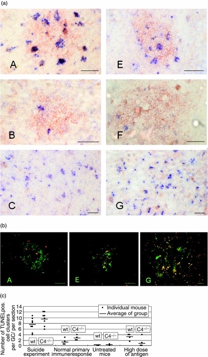
Deficiencies in early complement components are associated with the development of systemic lupus erythematosus (SLE) and therefore early complement components have been proposed to influence B lymphocyte activation and tolerance induction. A defect in apoptosis is a potential mechanism for breaking of peripheral B cell tolerance, and we hypothesized that the lack of the early complement component C4 could initiate autoimmunity through a defect in peripheral B lymphocyte apoptosis. Previous studies have shown that injection of a high dose of soluble antigen, during an established primary immune response, induces massive apoptotic death in germinal centre B cells. Here, we tested if the antigen-induced apoptosis within germinal centres is influenced by early complement components by comparing complement C4-deficient mice with C57BL/6 wild-type mice. We demonstrate that after the application of a high dose of soluble antigen in wild-type mice, antibody levels declined temporarily but were restored almost completely after a week. However, after antigen-induced apoptosis, B cell memory was severely limited. Interestingly, no difference was observed between wild-type and complement C4-deficient animals in the number of apoptotic cells, restoration of antibody levels and memory response.
Figures


Similar articles
-
Antigen localization within the splenic marginal zone restores humoral immune response and IgG class switch in complement C4-deficient mice.Int Immunol. 2004 Dec;16(12):1685-90. doi: 10.1093/intimm/dxh159. Epub 2004 Oct 11. Int Immunol. 2004. PMID: 15477230
-
Soluble antigen can cause enhanced apoptosis of germinal-centre B cells.Nature. 1995 May 25;375(6529):331-4. doi: 10.1038/375331a0. Nature. 1995. PMID: 7753199
-
Elevated levels of endogenous apoptotic DNA and IFN-alpha in complement C4-deficient mice: implications for induction of systemic lupus erythematosus.Eur J Immunol. 2007 Jun;37(6):1702-9. doi: 10.1002/eji.200636719. Eur J Immunol. 2007. PMID: 17506029
-
The association between systemic lupus erythematosus and deficiencies of the complement system.Cell Mol Biol (Noisy-le-grand). 2002 May;48(3):237-45. Cell Mol Biol (Noisy-le-grand). 2002. PMID: 12030427 Review.
-
B cell memory and the role of apoptosis in its formation.Mol Immunol. 2011 Jun;48(11):1301-6. doi: 10.1016/j.molimm.2010.10.026. Epub 2010 Dec 7. Mol Immunol. 2011. PMID: 21144588 Review.
References
-
- Pepys MB. Role of complement in induction of the allergic response. Nat New Biol. 1972;237:157–9. - PubMed
-
- Fischer MB, Ma M, Goerg S, et al. Regulation of the B cell response to T-dependent antigens by classical pathway complement. J Immunol. 1996;157:549–56. - PubMed
-
- Atkinson JP. Complement deficiency: predisposing factor to autoimmune syndromes. Clin Exp Rheumatol. 1989;7(Suppl. 3):S95–101. - PubMed
-
- Prodeus AP, Goerg S, Shen LM, et al A. critical role for complement in maintenance of self-tolerance. Immunity. 1998;9:721–31. - PubMed
-
- Botto M, Dell'Agnola C, Bygrave AE, et al. Homozygous C1q deficiency causes glomerulonephritis associated with multiple apoptotic bodies. Nat Genet. 1998;19:56–9. - PubMed
Publication types
MeSH terms
Substances
LinkOut - more resources
Full Text Sources
Medical
Molecular Biology Databases
Miscellaneous
