Review
. 2007 Oct;98(10):1528-35.
doi: 10.1111/j.1349-7006.2007.00567.x. Epub 2007 Jul 23.

PolyADP-ribosylation and cancer

Affiliations
Review

PolyADP-ribosylation and cancer

Masanao Miwa et al. Cancer Sci. 2007 Oct.

Abstract

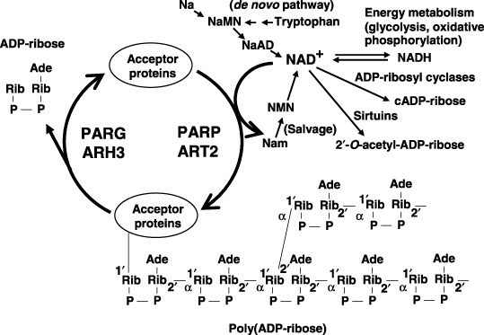
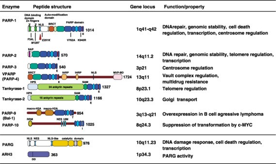
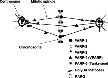
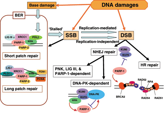
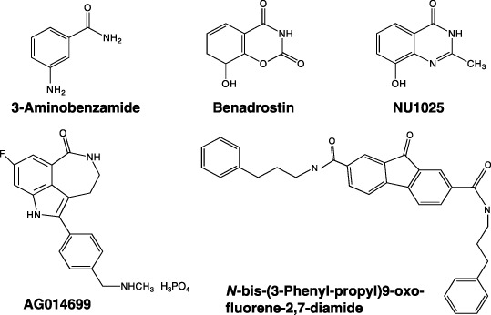
The polyADP-ribosylation reaction results in a unique post-translational modification involved in various cellular processes and conditions, including DNA repair, transcriptional control, genomic stability, cell death and transformation. The existence of 17 members of the poly(ADP-ribose) polymerase (PARP) family has so far been documented, with overlapping functional consequences. PARP-1 is known to be involved in DNA base excision repair and this explains the susceptibility spectrum of PARP-1 knockout animals to genotoxic carcinogens. The fact that centrosome amplification is induced by a non-genotoxic inhibitor of PARP and in PARP-1 knockout mouse cells, is in line with aneuploidy, which is frequent in cancers. Genetically engineered animal models have revealed that PARP-1 and VPARP impact carcinogenesis. Furthermore, accumulating experimental evidence supports the utility of PARP and PARG inhibitors in cancer therapy and several clinical trials are now ongoing. Increasing NAD(+) levels by pharmacological supplementation with niacin has also been found to exert preventive effects against cancer. In the present review, recent research progress on polyADP-ribosylation related to neoplasia is summarized and discussed.


Figures

Figure 1
Figure 1
PolyADP‐ribosylation and related reactions. ARH3, ADP‐ribose‐(arginine) protein hydrolase 3; ART2, monoADP‐ribosyl transferase 2; Na, nicotinic acid; NaAD, nicotinic acid adenine dinucleotide; NAD+ (βNAD+), nicotinamide adenine dinucleotide; Nam, nicotinamide; NaMN, nicotinic acid mononucleotide; NMN, nicotinamide mononucleotide; PARG, poly(ADP‐ribose) glycohydrolase; PARP, poly(ADP‐ribose) polymerases.
Figure 2
Figure 2
Poly(ADP‐ribose) polymerases (PARP) family proteins relating to carcinogenesis, poly(ADP‐ribose) glycohydrolase (PARG) and ADP‐ribose‐(arginine) protein hydrolase 3 (ARH3). Peptide structures, domains, motifs, gene loci and functions/properties are shown. For PARP‐1, positions of amino acids where single nucleotide polymorphisms (SNP) and mutations (italic) are shown. The caspase cleavage site is shown by the triangle. ARH, ADP‐ribosyl protein hydrolase; BRCT, BRCA1 C‐terminus; HPS, homopolymeric runs of His, Pro, and Ser; IHRP, inter‐α‐trypsin inhibitor family heavy chain‐related protein motif; MVP‐BD, major vault protein binding motif; NES, nuclear export signal; NLS, nuclear localization signal; RRM, RNA recognition motif; SAP, SAF‐A/B, acinus, and PIAS motif; SAM, a sterile α motif; UIM, ubiquitin‐interacting motif. The critical amino acid residue in the PARP domain of PARP‐1 and the residues at the corresponding position for each PARP family protein are shown. The DD residues indicated in ARH3 are critical residues for PARG activity.
Figure 3
Figure 3
Localization of poly(ADP‐ribose) polymerase (PARP) family members, poly(ADP‐ribose) glycohydrolase (PARG) and poly(ADP‐ribose) at the mitotic apparatus.
Figure 4
Figure 4
Model for involvement of poly(ADP‐ribose) polymerase (PARP) in DNA repair. DNA damage directly produces base damage, single‐strand breaks (SSB) and double‐strand breaks (DSB). SSB may be left unligated accidentally in the process of base excision repair (BER). SSB are possibly converted to DSB in a replication‐mediated manner or replication‐independently. Involvement of PARP in short‐patch and long‐patch repairs of BER, non‐homologous end‐joining (NHEJ) repair, and homologous recombination (HR) repair is shown. FEN1, flap endonuclease I; LIG, DNA ligase; PCNA, proliferating cell nuclear antigen; PNK, polynucleotide kinase; POL, DNA polymerase.
Figure 5
Figure 5
Developed poly(ADP‐ribose) polymerase (PARP) and poly(ADP‐ribose) glycohydrolase (PARG) inhibitors. Structures of PARP inhibitors, 3‐aminobenzamide,( 65 ) benadrostin,( 2 ) NU1025, AG014699,( 67 ) and PARG inhibitor N‐bis(3‐phenyl‐propyl)9‐oxo‐fluorene‐2,7‐diamide( 70 ) are shown.

References

    1. Sugimura T. Poly (adenosine diphosphate ribose). Prog Nucl Acid Res Mol Biol 1973; 13: 127–51. - PubMed
    1. Masutani M, Nakagama H, Sugimura T. Poly(ADP‐ribosyl)ation in relation to cancer and autoimmune disease. Cell Mol Life Sci 2005; 62: 769–83. - PMC - PubMed
    1. Miwa M, Kanai M, Uchida M, Uchida K, Hanai S. Roles of poly(ADP‐ribose) metabolism in the regulation of centrosome duplication and in the maintenance of neuronal integrity. In: Buerkle A, ed. Poly(ADP‐Ribosyl)ation. Georgetown, Texas: Landes Bioscience, 2006: 51–60
    1. Miwa M, Saikawa N, Yamaizumi Z, Nishimura S, Sugimura T. Structure of poly(adenosine diphosphate ribose): identification of 2′‐[1″‐ribosyl‐2″‐(or 3″‐) (1″′‐ribosyl) ]adenosine‐5′,5″,5″′‐tris (phosphate) as a branch linkage. Proc Natl Acad Sci USA 1979; 76: 595–9. - PMC - PubMed
    1. Althaus FR, Richter C. ADP‐ribosylation of proteins. Enzymology and biological significance. Mol Biol Biochem Biophys 1987; 37: 1–237. - PubMed

Publication types

Substances