Review
. 2007 Oct;98(10):1512-20.
doi: 10.1111/j.1349-7006.2007.00550.x. Epub 2007 Jul 23.

Signaling networks guiding epithelial-mesenchymal transitions during embryogenesis and cancer progression

Affiliations
Review

Signaling networks guiding epithelial-mesenchymal transitions during embryogenesis and cancer progression

Aristidis Moustakas et al. Cancer Sci. 2007 Oct.

Abstract

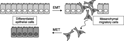
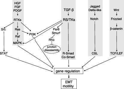
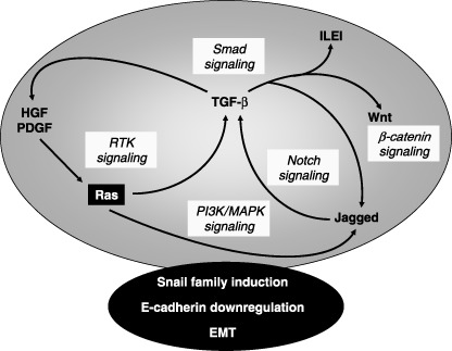
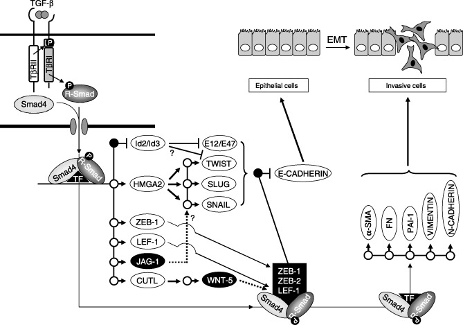
Epithelial-mesenchymal transition (EMT) describes the differentiation switch between polarized epithelial cells and contractile and motile mesenchymal cells, and facilitates cell movements and generation of new tissue types during embryogenesis. Many secreted polypeptides are implicated in the EMT process and their corresponding intracellular transduction pathways form highly interconnected networks. Transforming growth factor-beta, Wnt, Notch and growth factors acting through tyrosine kinase receptors induce EMT and often act in a sequential manner. Such growth factors orchestrate the concerted regulation of an elaborate gene program and a complex protein network, needed for establishment of new mesenchymal phenotypes after disassembly of the main elements of epithelial architecture, such as desmosomes, as well as tight, adherens and gap junctions. EMT of tumor cells occurs during cancer progression and possibly generates cell types of the tumor stroma, such as cancer-associated myofibroblasts. EMT contributes to new tumor cell properties required for invasiveness and vascular intravasation during metastasis. Here we present some of the current mechanisms that mediate the process of EMT and discuss their relevance to cancer progression.


Figures

Figure 1
Figure 1
Epithelial–mesenchymal transition (EMT) and mesenchymal–epithelial transition (MET). Cells in an epithelial sheet undergo EMT, generating motile mesenchymal derivatives (darker color/striped nuclei). Mesenchymal cells undergo MET, generating epithelial derivatives.
Figure 2
Figure 2
Major signal transduction pathways that induce epithelial–mesenchymal transition (EMT). Hepatocyte growth factor (HGF), fibroblast growth factor (FGF) and platelet‐derived growth factor (PDGF) signal via receptor tyrosine kinases (RTK) towards the central Ras‐Raf‐MAPK pathway or towards the PI3K pathway and the Src‐STAT pathway. Transforming growth factor (TGF)‐β signals via receptor serine/threonine kinases (RS/TK) towards the central R‐Smad/Co‐Smad pathway or towards the PI3K and MAPK pathways. Alternatively, the TGF‐β receptor signals towards the polarity protein Par6, thus recruiting the ubiquitin ligase Smurf, which degrades Rho and leads to disassembly of tight junctions in epithelial cells. Jagged and Delta‐like ligands signal via Notch receptors towards the transcription factor CSL. Wnt ligands signal via Frizzled receptors towards β‐catenin and the transcription factors LEF‐1/TCF. All these pathways modulate gene expression and lead to EMT and cell motility.
Figure 3
Figure 3
Autocrine/paracrine growth factor crosstalk during epithelial–mesenchymal transition (EMT). Autocrine or paracrine loops of growth factors induce expression and secretion of one another. Ras in the black box represents its oncogenic form that often is required in order to stimulate a robust autocrine crosstalk. The various signaling pathways involved are highlighted in light gray. The integrated signaling network (large gray ellipse) coordinates the action of transcription factors (such as Snail family members), which down‐regulate E‐cadherin among other gene responses in order to establish EMT.
Figure 4
Figure 4
The transcriptional program of transforming growth factor (TGF)‐β that elicits epithelial–mesenchymal transition (EMT). TGF‐β activates Smad complexes that act together with other transcription factors (TF) and induce expression of several other transcription factors. Gene targets are shown as circles. Black circles represent gene repression, while white circles represent gene induction. Id2 and Id3 inhibit E‐box‐binding proteins such as E12/E47 and possibly Twist (?). HMGA2 induces further expression of Twist, Slug or Snail. All these transcriptional repressors down‐regulate E‐cadherin expression. ZEB‐1 and LEF‐1 are transcriptionally induced and together with ZEB‐2 participate in transcriptional complexes with Smads, and repress the E‐cadherin gene. Jagged‐1 (JAG‐1) activates the Notch pathway (dotted arrow), which possibly induces expression of Snail (?). CUTL induces expression of Wnt‐5, which activates the β‐catenin pathway (dotted arrow), thus mobilizing transcription factor LEF‐1. Finally, Smad complexes induce expression of α‐smooth muscle actin (α‐SMA), fibronectin (FN), PAI‐1, vimentin and N‐cadherin, which contribute to the establishment of the motile, mesenchymal cell.

Similar articles

Cited by

References

    1. Hay ED. The mesenchymal cell, its role in the embryo, and the remarkable signaling mechanisms that create it. Dev Dyn 2005; 233: 706–20. - PubMed
    1. Huber MA, Kraut N, Beug H. Molecular requirements for epithelial‐mesenchymal transition during tumor progression. Curr Opin Cell Biol 2005; 17: 548–58. - PubMed
    1. Radisky DC, Kenny PA, Bissell MJ. Fibrosis and cancer: Do myofibroblasts come also from epithelial cells via EMT? J Cell Biochem; 101: 830–9. - PMC - PubMed
    1. Thiery J‐P, Sleeman JP. Complex networks orchestrate epithelial‐mesenchymal transitions. Nat Rev Mol Cell Biol 2006; 7: 131–42. - PubMed
    1. Zeisberg M, Kalluri R. The role of epithelial‐to‐mesenchymal transition in renal fibrosis. J Mol Med 2004; 82: 175–81. - PubMed

Publication types