

Immunohistochemical features of a papillary squamous cell carcinoma of the endometrium with transitional cell differentiation
- PMID: 17645802
- PMCID: PMC1947947
- DOI: 10.1186/1746-1596-2-26
Immunohistochemical features of a papillary squamous cell carcinoma of the endometrium with transitional cell differentiation
Abstract
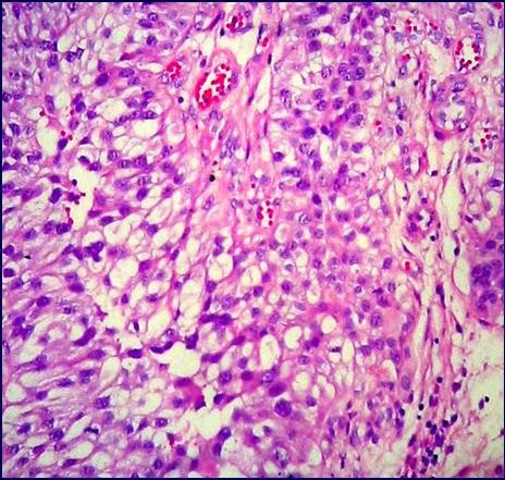
An 84-year-old woman underwent hysterectomy due to a friable endometrial mass infiltrating almost half way through the myometrial wall. The tumor consisted of papillary structures with thin fibrovascular cores covered by several layers of pleomorphic cells. The deeply located neoplastic cells were ovoid with a pale eosinophilic cytoplasm resembling urothelial cells. A diagnosis of papillary squamous cell carcinoma of the endometrium with transitional cell differentiation was made. Although she recovered well after surgery, she died one year later because of disseminated disease. In an attempt to obtain new insights into the physiopathology of this very rare tumor, an immunohistochemical panel with 32 markers was performed. The neoplastic cells were positive for cytokeratin 5, vimentin, p63, p21, VEGF, Ki67, BAG1, and bcl-2. The expression of BAG-1 and bcl-2 may suggest that anti-apoptotic stimuli are preponderant in this neoplasm.
Figures


Similar articles
-
Transitional cell carcinoma of the endometrium and endometrial carcinoma with transitional cell differentiation: a clinicopathologic study of 5 cases and review of the literature.Hum Pathol. 2008 Nov;39(11):1606-13. doi: 10.1016/j.humpath.2008.03.005. Epub 2008 Jul 11. Hum Pathol. 2008. PMID: 18620731 Review.
-
Plasmacytoid urothelial carcinoma of the urinary bladder: a case report and immunohistochemical study.Pathol Res Pract. 2009;205(3):189-94. doi: 10.1016/j.prp.2008.09.004. Epub 2008 Nov 28. Pathol Res Pract. 2009. PMID: 19041193
-
Invasive urothelial carcinoma with chordoid features: a report of 12 distinct cases characterized by prominent myxoid stroma and cordlike epithelial architecture.Am J Surg Pathol. 2009 Aug;33(8):1213-9. doi: 10.1097/PAS.0b013e3181a8ffbe. Am J Surg Pathol. 2009. PMID: 19542871
-
Plasmacytoid transitional cell carcinoma of urinary bladder: a clinicopathologic study of 9 cases.Am J Surg Pathol. 2008 May;32(5):752-7. doi: 10.1097/PAS.0b013e318159af9e. Am J Surg Pathol. 2008. PMID: 18379419
-
How to combine the molecular profile with the clinicopathological profile of urothelial neoplastic lesions.Scand J Urol Nephrol Suppl. 2008 Sep;(218):175-84. doi: 10.1080/03008880802291873. Scand J Urol Nephrol Suppl. 2008. PMID: 18815932 Review.
Cited by
-
Primary endometrioid adenocarcinoma of the cervix with widespread squamous metaplasia--a potential diagnostic pitfall.Diagn Pathol. 2007 Oct 25;2:40. doi: 10.1186/1746-1596-2-40. Diagn Pathol. 2007. PMID: 17961245 Free PMC article.
-
Case report: Clinicopathological characteristic of two cases of primary endometrial squamous cell carcinoma and review of the literature.Front Oncol. 2024 Aug 26;14:1415816. doi: 10.3389/fonc.2024.1415816. eCollection 2024. Front Oncol. 2024. PMID: 39252944 Free PMC article.
-
Endometrioid adenocarcinoma with simultaneous endocervical and intestinal-type mucinous differentiation: report of a rare phenomenon and the immunohistochemical profile.Diagn Pathol. 2013 Aug 2;8:128. doi: 10.1186/1746-1596-8-128. Diagn Pathol. 2013. PMID: 23915109 Free PMC article.
-
Expression of BAG-1 and PARP-1 in precursor lesions and invasive cervical cancer associated with human papillomavirus (HPV).Pathol Oncol Res. 2012 Oct;18(4):929-37. doi: 10.1007/s12253-012-9523-y. Epub 2012 Mar 28. Pathol Oncol Res. 2012. PMID: 22454210
-
Transitional cell carcinoma of endometrium: a case report of rare pure form.J Contemp Brachytherapy. 2018 Oct;10(5):483-485. doi: 10.5114/jcb.2018.79411. Epub 2018 Oct 31. J Contemp Brachytherapy. 2018. PMID: 30479627 Free PMC article.
References
-
- Rodolakis A, Papaspyrou I, Sotiropoulou M, Markaki S, Michalas S. Primary squamous cell carcinoma of the endometrium. A report of 3 cases. Eur J Gynaecol Oncol. 2001;22:143–146. - PubMed
LinkOut - more resources
Full Text Sources
Research Materials
