. 2007 Sep 21;282(38):27562-71.
doi: 10.1074/jbc.M700340200. Epub 2007 Jul 23.

NeuA sialic acid O-acetylesterase activity modulates O-acetylation of capsular polysaccharide in group B Streptococcus

Affiliations

NeuA sialic acid O-acetylesterase activity modulates O-acetylation of capsular polysaccharide in group B Streptococcus

Amanda L Lewis et al. J Biol Chem. .

Abstract

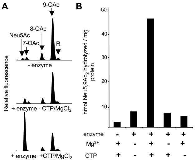
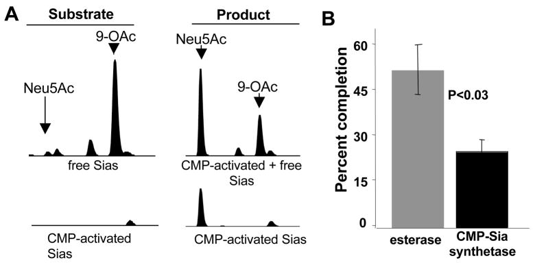
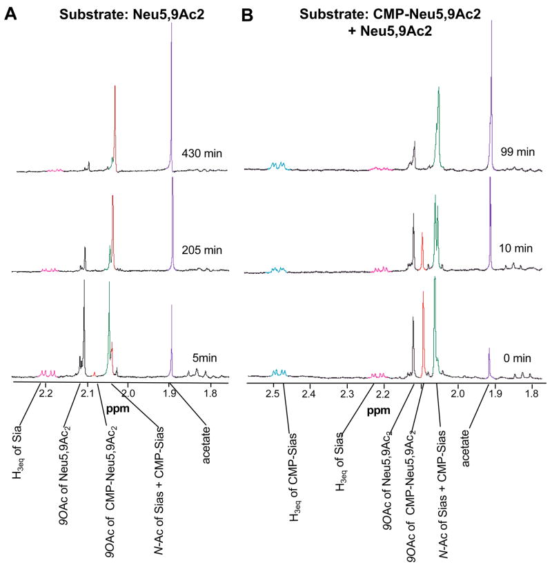
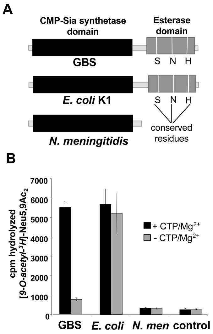
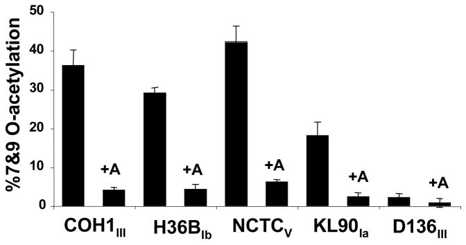
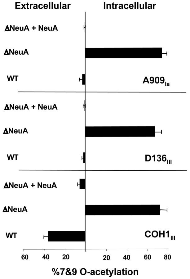
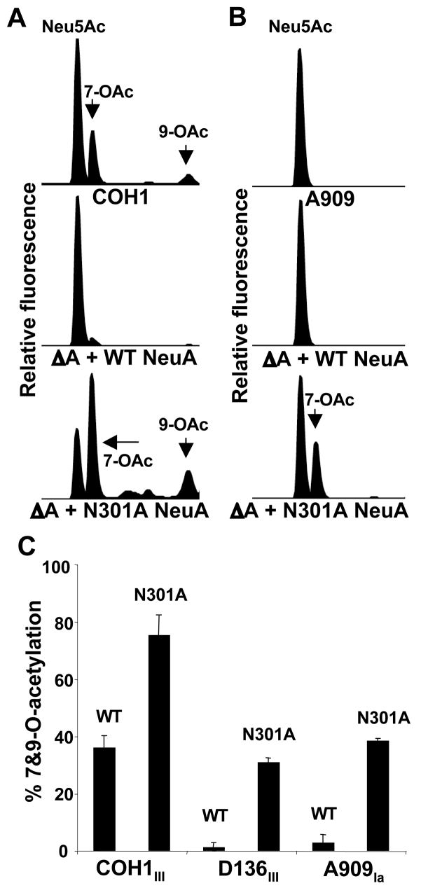
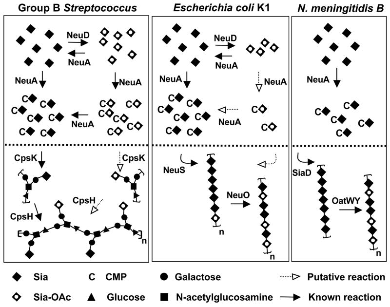
Group B Streptococcus (GBS) is a common cause of neonatal sepsis and meningitis. A major GBS virulence determinant is its sialic acid (Sia)-capped capsular polysaccharide. Recently, we discovered the presence and genetic basis of capsular Sia O-acetylation in GBS. We now characterize a GBS Sia O-acetylesterase that modulates the degree of GBS surface O-acetylation. The GBS Sia O-acetylesterase operates cooperatively with the GBS CMP-Sia synthetase, both part of a single polypeptide encoded by the neuA gene. NeuA de-O-acetylation of free 9-O-acetyl-N-acetylneuraminic acid (Neu5,9Ac(2)) was enhanced by CTP and Mg(2+), the substrate and co-factor, respectively, of the N-terminal GBS CMP-Sia synthetase domain. In contrast, the homologous bifunctional NeuA esterase from Escherichia coli K1 did not display cofactor dependence. Further analyses showed that in vitro, GBS NeuA can operate via two alternate enzymatic pathways: de-O-acetylation of Neu5,9Ac(2) followed by CMP activation of Neu5Ac or activation of Neu5,9Ac(2) followed by de-O-acetylation of CMP-Neu5,9Ac(2). Consistent with in vitro esterase assays, genetic deletion of GBS neuA led to accumulation of intracellular O-acetylated Sias, and overexpression of GBS NeuA reduced O-acetylation of Sias on the bacterial surface. Site-directed mutagenesis of conserved asparagine residue 301 abolished esterase activity but preserved CMP-Sia synthetase activity, as evidenced by hyper-O-acetylation of capsular polysaccharide Sias on GBS expressing only the N301A NeuA allele. These studies demonstrate a novel mechanism regulating the extent of capsular Sia O-acetylation in intact bacteria and provide a genetic strategy for manipulating GBS O-acetylation in order to explore the role of this modification in GBS pathogenesis and immunogenicity.


Figures

Figure 1
Figure 1. Purified GBS NeuA hydrolyzes 9-O-acetylated Neu5Ac in a CTP- and Mg2+-dependent manner
His-tagged GBS NeuA was purified and 9-O-acetylated Neu5Ac (“Neu5,9Ac2” or “9-OAc”) was synthesized using a chemoenzymatic approach as described in Experimental Procedures. Enzyme assays were performed in 100 mM Tris pH 7.5 with 1μM enzyme and 8 μM substrate and allowed to proceed for 40 min followed by DMB derivatization and HPLC resolution of the 9-O-acetylated substrate “9-OAc,” and non-O-acetylated product “Neu5Ac”. “R” is a reagent peak of unknown identity. A. HPLC profiles of reactions performed in the presence or absence of enzyme or CMP-Sia synthetase co-factors (CTP and Mg2+) B. Quantitation of Sia-O-acetyl hydrolysis by integration of Neu5Ac peak. Optimal NeuA esterase activity requires the presence of both CTP (5 mM) and MgCl2 (20 mM).
Figure 2
Figure 2. GBS NeuA can directly de-O-acetylate Neu5,9Ac2
NeuA esterase and CMP-Sia synthetase activities were monitored to test the hypothesis that CTP/Mg2+ dependence is due to a processive mechanism. A. NeuA enzyme assays were performed in 100 mM Tris pH 7.5 with (right) or without (left) 2 μM enzyme in the presence of 8 μM substrate and allowed to proceed for 30 min. The CMP-Sia bond is labile under the acidic conditions of DMB derivatization, thereby resulting in combined detection of free and CMP-bound Sias (top). Half of each reaction was treated with 80 mM freshly prepared NaBH4 to destroy free Sias and detect only CMP-activated Sias (bottom). See Figure 1 legend for complete description of peaks. B. Percent completion of CMP-synthetase and esterase reactions in A. Sia-O-acetyl hydrolysis was measured as a percentage of total Sia substrate in the reaction, using integration of Neu5Ac peaks in samples +/− enzyme without NaBH4 treatment. CMP-Sia activation was likewise measured by integration of peaks in the NaBH4-treated samples. While 52% of the Neu5,9Ac2 substrate was de-O-acetylated, only 25% of the free Sias were CMP-activated. These data are representative of 2 independent experiments.
Figure 3
Figure 3. CMP-Neu5,9Ac2 is a direct substrate of the NeuA esterase and is de-O-acetylated more rapidly than free Neu5,9Ac2
NeuA reactions were monitored by 600 MHz 1H NMR. Experiments were carried out at 37 °C in a total volume of 0.7 mL. Assays included 2.5 μM GBS NeuA with 2.5 mM substrate, CTP and MgCl2 in 20 mM deuterated phosphate buffer pH 7.5. About 5 min elapsed between the addition of the enzyme, and the acquisition of the first time point (defined as t = 0 min). NMR spectra were acquired every 2 minutes. Data shown are representative of the trends observed throughout the reaction. A. Neu5,9Ac2 alone was used as substrate. B. Enzymatically synthesized CMP-Neu5,9Ac2 was used as substrate, but contained a significant amount of free Neu5,9Ac2 allowing internal comparison.
Figure 4
Figure 4. Divergence of co-factor dependence between the homologous bi-functional NeuA enzymes of GBS and E. coli
A. Schematic representation of NeuA homologs in GBS, E. coli K1 and N. meningitidis. B. Radioligand hydrolysis assay comparing requirements of NeuA esterase activity between the three homologs. Reactions were performed as described in Figure 2 with (or without) 4 μM enzyme in the presence or absence of 5 mM CTP and 20 mM MgCl2. Bars represent standard deviation of 2 experiments performed on separate days.
Figure 5
Figure 5. Over-expression of GBS NeuA decreases Sia-O-acetylation on GBS strains of multiple serotypes
WT GBS strains were transformed with an expression construct encoding the full-length NeuA enzyme “+A” and O-acetylation was compared to the parent strain. Total cellular Sias were isolated by mild acid hydrolysis and fluorescently derivatized with DMB as described in Experimental Procedures. Derivatized Sias were resolved by reverse phase HPLC and percent O-acetylation determined by software-assisted integration of HPLC peaks corresponding to O-acetylated and non-O-acetylated Neu5Ac peaks. HPLC profiles of the COH1 strain and corresponding NeuA-overexpressing strain were published previously (17).
Figure 6
Figure 6. Deletion of GBS NeuA increases intracellular Sia-O-acetylation in both “high-OAc” and “low-OAc” strains
Elimination of NeuA (both CMP-Sia synthestase and esterase domains) in different GBS strains was accomplished by precise allelic replacement of the neuA gene with chloramphenicol acetyltransferase (cat) to produce the ΔNeuA or “ΔA” strains as described in Experimental Procedures. Mutant strains were complemented by plasmid-based expression of NeuA (“+A”) as in Figure 4. Intracellular and extracellular Sias were separated as previously described (17) and percent O-acetylation was determined by DMB-HPLC analysis. HPLC profiles of the wild-type COH1 and corresponding ΔNeuA strain were published previously (17). Bars represent standard deviation of 2 or more independent experiments.
Figure 7
Figure 7. Active site mutagenesis of the NeuA esterase increases capsular O-acetylation
Site-directed mutagenesis of putative esterase active site residue Asn301 was performed using the WT NeuA expression plasmid as template. “WT NeuA” and mutated “N301A NeuA” constructs were then used to complement all three GBS NeuA-deficient “ΔA” strains, followed by DMB-HPLC analysis of sialidase-released (capsular) Sias. Raw HPLC data for “WT”, “ΔA + WT NeuA”, and “ΔA + N301A NeuA” strains in the A. COH1 (high-OAc) and B. A909 (low-OAc) GBS backgrounds. C. Percent O-acetylation of N301A-complemented NeuA-deficient “ΔA + N301A NeuA” strains is substantially higher than the respective isogenic WT NeuA-complemented strains. Further experiments employing separation of intracellular and extracellular Sias from each strain validated the lack of intracellular Sia accumulation and capsular hyper-O-acetylation of the “ΔA + N301A NeuA” strains (data not shown). Bars represent standard deviation of 2 or more independent experiments.
Figure 8
Figure 8. NeuA esterase activity in the context of capsule biosynthetic evolution in three prominent Sia-decorated pathogens GBS, E. coli K1, and serogroup B N. meningitidis.
Bacterial Sia biosynthesis can be divided into three main parts: 1) Sia biosynthesis and degradation, 2) intracellular Sia O-acetylation and de-O-acetylation, and 3) polysaccharide biosynthesis, polymerization, and O-acetylation. For simplicity, only 2) and 3) are shown (above and below the dotted line respectively). With respect to 1), these Sia-expressing pathogens use a homologous 3-enzyme pathway to generate N-acetylmannosamine (ManNAc) by epimerization of UDP-N-acetylglucosamine, condense phosphoenolpyruvate with ManNAc to generate N-acetylneuraminic acid (Neu5Ac), then activate Neu5Ac by transfer of the anomeric oxygen to the α-phosphate of CTP. Only E. coli possesses Sia degradation machinery. In GBS, cpsK and cpsH encode the sialyltransferase and repeating unit polymerase respectively. Intracellular Sia O-acetylation and de-O-acetylation is limited to GBS and E. coli, both of which possess the NeuD Sia O-acetyltransferase and NeuA Sia-O-acetyl esterase. The N. meningitidis pathway is more stream-lined in appearance due to the lack of an endogenous O-acetylation/de-O-acetylation cycle. Despite the pronounced difference between N. meningitidis (serogroup B) and E. coli K1 in intracellular O-acetylation cycling, these pathogens share an identical poly-Sia antigen made up of α2–8-linked Neu5Ac residues (polymerized by SiaD and NeuS respectively) that can be O-acetylated after capsule polymerization (by OatWY or NeuO respectively). See text for further discussion. Model is based on references (, , , –61).

Similar articles

Cited by

References

    1. Angata T, Varki A. Chem Rev. 2002;102:439–469. - PubMed
    1. Gagneux P, Varki A. Glycobiology. 1999;9:747–755. - PubMed
    1. Vimr E, Lichtensteiger C. Trends Microbiol. 2002;10:254–257. - PubMed
    1. Edwards MS, Kasper DL, Jennings HJ, Baker CJ, Nicholson W. J Immunol. 1982;128:1278–1283. - PubMed
    1. Marques MB, Kasper DL, Pangburn MK, Wessels MR. Infect Immun. 1992;60:3986–3993. - PMC - PubMed

Publication types

MeSH terms

LinkOut - more resources