. 2007 Jul 30;178(3):399-410.
doi: 10.1083/jcb.200702167. Epub 2007 Jul 23.

Yeast peroxisomes multiply by growth and division

Affiliations

Yeast peroxisomes multiply by growth and division

Alison M Motley et al. J Cell Biol. .

Abstract

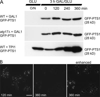
Peroxisomes can arise de novo from the endoplasmic reticulum (ER) via a maturation process. Peroxisomes can also multiply by fission. We have investigated how these modes of multiplication contribute to peroxisome numbers in Saccharomyces cerevisiae and the role of the dynamin-related proteins (Drps) in these processes. We have developed pulse-chase and mating assays to follow the fate of existing peroxisomes, de novo-formed peroxisomes, and ER-derived preperoxisomal structures. We find that in wild-type (WT) cells, peroxisomes multiply by fission and do not form de novo. A marker for the maturation pathway, Pex3-GFP, is delivered from the ER to existing peroxisomes. Strikingly, cells lacking peroxisomes as a result of a segregation defect do form peroxisomes de novo. This process is slower than peroxisome multiplication in WT cells and is Drp independent. In contrast, peroxisome fission is Drp dependent. Our results show that peroxisomes multiply by growth and division under our assay conditions. We conclude that the ER to peroxisome pathway functions to supply existing peroxisomes with essential membrane constituents.


Figures

Figure 1.
Figure 1.
Models of peroxisome formation and multiplication. The first two models propose that peroxisomes form de novo from the ER. ER-derived membrane structures (preperoxisomes) mature into peroxisomes that import matrix proteins (black). The role of Drps has been suggested to be either at the ER membrane (model 1) or at a later stage in the maturation pathway (model 2). The third model proposes that peroxisomes multiply by fission of existing peroxisomes (black) and that the ER provides lipids and some membrane proteins in the form of a preperoxisomal structure (gray) that fuses with existing peroxisomes. Drps have been proposed to be required for the fission of peroxisomes.
Figure 2.
Figure 2.
Pulse-chase analysis of GFP-PTS1–labeled peroxisomes. (A) Western blot analysis of a GFP-PTS1 pulse-chase experiment. Cells transformed with GFP-PTS1–expressing plasmids under the control of either the inducible GAL1 promoter or the constitutive TPI1 promoter were grown overnight on glucose, shifted to galactose medium for 3 h (pulse), and subsequently transferred to glucose medium (chase) for 6 h. Samples of equal culture volume (1 ml) were collected during the chase and analyzed at each time point. At t = 0, 1 ml contains 0.05 OD600 units. Whereas GAL1-regulated expression is shut off and the level of GFP-PTS1 remains constant, TPI1-regulated expression increases. For the overnight glucose-grown sample, 0.15 OD units were loaded. (B) Fluorescence micrograph of pulse-labeled cells at t = 120 min and t = 360 min (left and middle). The same exposure time was used without enhancement of the signal. Note the decrease in fluorescence intensity. (right) GFP signal is enhanced to illustrate that the number of fluorescent peroxisomes per cell remains constant. Bar, 5 μm.
Figure 3.
Figure 3.
Mating assay to study peroxisome dynamics and formation. (A) Peroxisomes do not fuse with each other. Peroxisomes were fluorescently pulse labeled with either GFP or HcRed in WT cells of opposite mating types by growth on galactose medium for 3 h followed by a chase on glucose medium for 2 h. Subsequently, cells were allowed to mate for 2 h (first to third rows) or 4 h (fourth row) before fixing. Cells were imaged at different stages of mating. No colocalization between GFP and HcRed was seen. (B) All peroxisomes are import competent. WT and pex3Δ cells were pulse labeled with GFP-PTS1 and HcRed-PTS1, respectively, chased for 2 h, and mated and processed for imaging as in A. Upon cytoplasmic mixing, all GFP-labeled peroxisomes import HcRed-PTS1, although to a varying extent. No peroxisomes were seen that contained HcRed-PTS1 only. (C) De novo peroxisome formation upon mating is a slow process. pex3Δ cells constitutively expressing HcRed-PTS1 were mated with pex19Δ cells. From left to right, the panels show images of cells taken 3, 5, and 7 h after mating. (D) Quantitation of the rate of de novo peroxisome formation (see C). The number of mating cells containing peroxisomes was counted at each time point and expressed as a percentage of total mating cells containing HcRed-PTS1. For each time point, at least 100 mating cells were analyzed. Bars, 5 μm.
Figure 4.
Figure 4.
Peroxisomes are formed de novo only in the absence of preexisting peroxisomes. (A and D) WT (A) and inp2Δ (D) cells constitutively expressing HcRed-PTS1 and conditionally expressing GFP-PTS1 were pulse labeled for 3 h on galactose medium and chased for 2 h on glucose medium. Cells were then seeded thinly onto a glucose-containing agarose pad on a microscope slide and allowed to grow for 6–8 h before imaging, which is long enough to allow single budding cells to give rise to a colony. Any peroxisomes that are formed de novo after the shutdown of GFP-PTS1 expression will label with HcRed only. The level of GFP signal was enhanced relative to the level of RFP. (A) All cells in the WT colony contain peroxisomes that label with both GFP and HcRed. However, GFP and HcRed do not overlap completely because the colonies could not be fixed. (B) Analysis of WT cells grown in liquid culture after 6 h of chase. All HcRed-labeled peroxisomes also labeled with GFP (Fig. 4 B), indicating that all peroxisomes are derived from those present before GFP expression was shut down. (C) Mating assay to test for de novo–formed peroxisomes. GFP-PTS1 expression was induced for 3 h in WT cells followed by a 5-h chase on glucose. WT cells were then mated with pex3Δ cells expressing HcRed-PTS1. No red-only peroxisomes were detected, indicating that no peroxisomes are formed de novo during the 5-h chase. (D) In inp2Δ colonies, usually only one or two cells contained GFP, these cells comprising the original GFP-expressing cells from which the rest of the colony was derived. Approximately half of the cells in each colony contain peroxisomes that are labeled with HcRed-PTS1. (E) Bar graph showing the proportion of cells (>150 cells were counted) that had formed peroxisomes de novo 6–8 h after seeding onto agarose (see A). Colonies were examined for the presence of cells with no peroxisomes, red/green (preexisting) peroxisomes, or exclusively red-only (de novo formed) peroxisomes. Bars, 5 μm.
Figure 5.
Figure 5.
Pex3-GFP is targeted to existing peroxisomes. (A) Newly synthesized Pex3-GFP associates with all peroxisomes present in WT cells. Pex3-GFP expression was induced for 3 h in cells constitutively expressing HcRed-PTS1. Pex3-GFP colocalizes completely with peroxisomes in cells with low Pex3-GFP expression. In some cells with higher expression, faint additional Pex3-GFP punctae were observed. It is not clear whether these Pex3-GFP punctae are an early stage of de novo peroxisome formation induced by the overexpression of Pex3p or whether they are aggregates that will later be degraded. (B) Pex3-GFP trapped in the ER in pex19Δ cells is released upon mating with WT cells and associates with preexisting peroxisomes. pex19Δ cells pulse labeled with Pex3-GFP (3 h of galactose and 2 h of glucose) were mated with WT cells pulse labeled with HcRed-PTS1 (3 h of galactose and 2 h of glucose). Cells were fixed after 2 h. The Pex3-GFP signal that colocalizes with HcRed becomes stronger with time after mating. Bars, 5 μm.
Figure 6.
Figure 6.
Vps1p is required for fission of existing peroxisomes. Peroxisomes in vps1Δ/dnm1Δ cells were pulse labeled with GFP-PTS1 (3 h of galactose and 2 h of glucose). (A–C) pex3Δ (A), pex3Δ/vps1Δ (B), and pex3Δ/vps1Δ cells overexpressing Dnm1p (C) were pulse labeled with HcRed-PTS1 in the same way. Cells were mated for 2 h before fixing and imaging. After cell fusion and cytoplasmic mixing, HcRed-PTS1 is imported into the GFP-labeled peroxisomal structures (A–C), which, in the presence of Vps1p (A) or overexpressed Dnm1p (C), are divided into multiple peroxisomes. No fission occurs in the absence of Vps1p (B). (D) Time-lapse microscopy of fission after vps1Δ/dnm1Δ cells were mated with pex3Δ cells. Bars, 5 μm.
Figure 7.
Figure 7.
Drps are not required for the ER to peroxisome transport of Pex3-GFP or for de novo peroxisome formation. (A) Newly synthesized Pex3-GFP associates with the single peroxisomal structure present in vps1Δ/dnm1Δ cells. In cells labeled with HcRed-PTS1, the expression of Pex3-GFP was induced for 3 h. Most GFP colocalizes with peroxisomes, although some faint additional punctae were observed. (B and C) vps1Δ/dnm1Δ (B) and vps1Δ/dnm1Δ/inp2Δ (C) cells constitutively expressing HcRed-PTS1 and conditionally expressing GFP-PTS1 were pulse labeled for 3 h on galactose and chased for 2 h on glucose medium. Cells were then seeded onto a glucose-containing agarose pad on a microscope slide and allowed to grow for 8 h before imaging. In vps1Δ/dnm1Δ/inp2Δ colonies, peroxisomes form independently of Vps1p and Dnm1p. GFP labeled a single peroxisomal structure in a single cell in each colony, whereas approximately half of the cells contain multiple peroxisomes that label with HcRed-PTS1, which is indicative of de novo–formed peroxisomes. As expected, with high exposure time, a very faint cytoplasmic labeling is visible in some of the cells that lack peroxisomes (C, arrowhead; red-only panel). (D) Bar graph showing the proportion of cells (>150 cells were counted) that had formed peroxisomes de novo ∼8 h after seeding onto agarose (see B). Colonies were examined for the presence of cells with no peroxisomes, red/green (preexisting) peroxisomes, or exclusively red-only (de novo formed) peroxisomes. Bars, 5 μm.

References

    1. Cerveny, K.L., J.M. McCaffery, and R.E. Jensen. 2001. Division of mitochondria requires a novel DMN1-interacting protein, Net2p. Mol. Biol. Cell. 12:309–321. - PMC - PubMed
    1. Cheong H., T. Yorimitsu, F. Reggiori, J.E. Legakis, C.W. Wang, and D.J. Klionsky. 2005. Atg17 regulates the magnitude of the autophagic response. Mol. Biol. Cell 16:3438–3453. - PMC - PubMed
    1. Duttagupta, R., S. Vasudevan, C.J. Wilusz, and S.W. Peltz. 2003. A yeast homologue of Hsp70, Ssa1p, regulates turnover of the MFA2 transcript through its AU-rich 3′ untranslated region. Mol. Cell. Biol. 23:2623–2632. - PMC - PubMed
    1. Fagarasanu, A., M. Fagarasanu, G.A. Eitzen, J.D. Aitchison, and R.A. Rachubinski. 2006. The peroxisomal membrane protein Inp2p is the peroxisome-specific receptor for the myosin V motor Myo2p of Saccharomyces cerevisiae. Dev. Cell. 10:587–600. - PubMed
    1. Fagarasanu, M., A. Fagarasanu, Y.Y. Tam, J.D. Aitchison, and R.A. Rachubinski. 2005. Inp1p is a peroxisomal membrane protein required for peroxisome inheritance in Saccharomyces cerevisiae. J. Cell Biol. 169:765–775. - PMC - PubMed

Publication types

MeSH terms