. 2007 Aug 6;204(8):1945-57.
doi: 10.1084/jem.20070133. Epub 2007 Jul 23.

The role of the Runx transcription factors in thymocyte differentiation and in homeostasis of naive T cells

Affiliations

The role of the Runx transcription factors in thymocyte differentiation and in homeostasis of naive T cells

Takeshi Egawa et al. J Exp Med. .

Erratum in

  • J Exp Med. 2008 Aug 4;205(8):1939

Abstract

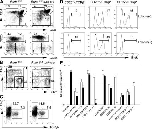
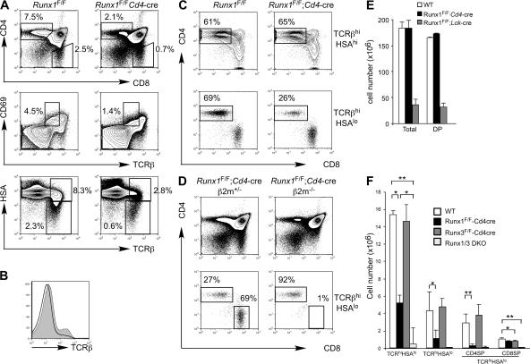
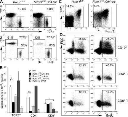
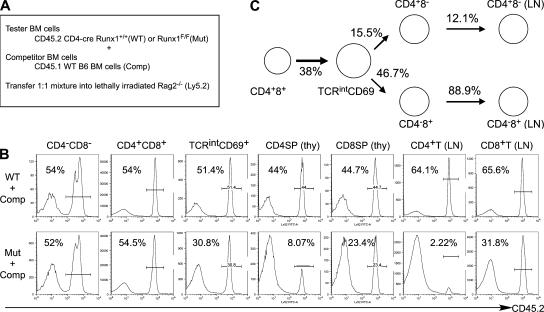
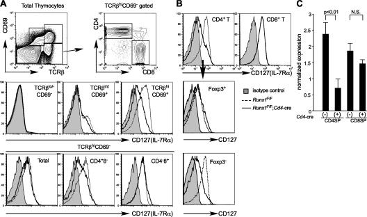
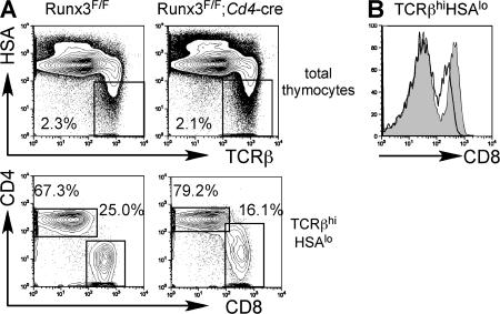
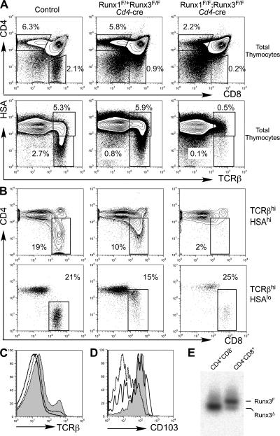
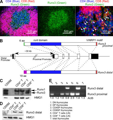
Members of the Runx family of transcriptional regulators are required for the appropriate expression of CD4 and CD8 at discrete stages of T cell development. The roles of these factors in other aspects of T cell development are unknown. We used a strategy to conditionally inactivate the genes encoding Runx1 or Runx3 at different stages of thymocyte development, demonstrating that Runx1 regulates the transitions of developing thymocytes from the CD4(-)CD8(-) double-negative stage to the CD4(+)CD8(+) double-positive (DP) stage and from the DP stage to the mature single-positive stage. Runx1 and Runx3 deficiencies caused marked reductions in mature thymocytes and T cells of the CD4(+) helper and CD8(+) cytotoxic T cell lineages, respectively. Runx1-deficient CD4(+) T cells had markedly reduced expression of the interleukin 7 receptor and exhibited shorter survival. In addition, inactivation of both Runx1 and Runx3 at the DP stages resulted in a severe block in development of CD8(+) mature thymocytes. These results indicate that Runx proteins have important roles at multiple stages of T cell development and in the homeostasis of mature T cells.


Figures

Figure 1.
Figure 1.
Runx1 requirement in the differentiation of DN thymocytes. (A) CD4 and CD8 expression in total thymocytes (top) and CD25 and CD44 expression in CD4lo/−CD8TCRβ thymocytes (bottom) from Runx1 F/F;Lck-cre mice and littermate Runx1 F/F control mice. Percentages of cells are shown in the indicated gates. (B and C) CD25 and icTCRβ (B) and icTCRβ and TCRγδ (C) in CD4lo/−CD8TCRβ thymocytes are shown. Gates used for population definition and frequencies of the gated population in CD4lo/−CD8TCRβ thymocytes are indicated. (D) Incorporation of BrdU by the thymocyte subpopulations defined in B. Numbers indicate the percentages of BrdU+ cells defined by the interval gates. (E) Absolute numbers of thymocytes in immature thymocyte subpopulations from Runx1 F/F;Lck-cre mice (closed bars) and littermate Runx1 F/F mice (open bars) are shown. Data were averaged from five mice with standard deviations.
Figure 2.
Figure 2.
Runx1 is required for positive selection and maturation of CD4SP thymocytes. (A) Expression of CD4, CD8, TCRβ, CD69, and HSA (CD24) in thymocytes from Runx1 F/F;Cd4-cre and littermate Runx1 F/F mice (top) is shown. Percentages of cells are shown in the indicated gates. (B) TCRβ expression of total thymocytes from Runx1 F/F;Cd4-cre (open histogram) and littermate Runx1 F/F (shaded histogram) mice. (C) CD4 and CD8 expression in thymocytes gated for TCRβhiHSAhi (top) and TCRβhiHSAlo (bottom) from Runx1 F/F;Cd4-cre and littermate Runx1 F/F mice. Percentages of CD4+CD8 thymocytes in the gated rectangles are shown. (D) Development of CD4SP and CD8SP thymocytes in Runx1 F/F;Cd4-cre and littermate Runx1 F/F;Cd4-cre; b2m −/− mice. The percentages of CD4SP and CD8SP mature thymocytes are shown. (E and F) The absolute numbers of total thymocytes and DP thymocytes (E) and mature thymocyte subpopulations (F) are shown as the mean and standard deviation from 4–10 mice with the indicated genotypes. Statistical differences were tested by the Student's t test. *, P < 0.05; **, P < 0.01.
Figure 3.
Figure 3.
Runx1 regulates homeostasis of CD4+ T cells in the periphery. (A) TCRβ and CD19 expression (top) and CD4 and CD8 expression in gated TCRβ+ cells (bottom). The percentages of populations gated in the rectangles are shown. (B) Numbers of TCRβ+ cells, CD4+ T cells, and CD8+ T cells from spleens are shown as the means and standard deviations from 4–10 mice of each indicated genotype. (C) Foxp3 and CD25 expression in CD4+ T cells. ­Peripheral lymphocytes were stained for TCRβ, CD4, CD25, and intracellular Foxp3, and Foxp3 and CD25 expression in TCRβ+CD4+CD8 cells is shown. The percentages of Foxp3+ cells gated by the rectangles are shown. (D) BrdU incorporation by peripheral lymphocytes in Runx1 F/F;Cd4-cre and littermate Runx1 F/F mice. Mice were administered BrdU for 72 h in drinking water, and percentages of BrdU+ cells in gated B cells, CD4+ T cells, and CD8+ T cells are shown. Data shown represent three experiments with similar results. Statistical differences were tested by the Student's t test. *, P < 0.05; **, P < 0.01.
Figure 4.
Figure 4.
Quantitative analysis of precursor activity of Runx1-deficient thymocytes by a competitive repopulation assay. (A) Strategy for generation of mixed bone chimeras with CD45.2+ bone marrow cells from Runx1 F/F;Cd4-cre (Mut) or littermate control Cd4-cre (WT) mice. CD45.2+ bone marrow cells mixed with wild-type CD45.2 (CD45.1; Comp) competitor bone marrow cells at the ratio of 1:1 were transferred into lethally irradiated Rag2−/− hosts. T cell development was analyzed 10–12 wk after transplantation. (B) Contribution of CD45.2+ bone marrow cells at different stages of T cell development. Reconstituted thymocytes or lymph node cells were analyzed for frequency of CD45.2+-expressing cells in populations defined by CD4, CD8, TCRβ, and CD69 expression. (C) Relative T cell precursor potential of Runx1-deficient thymocytes, as determined in the competitive repopulation assay. The values for T cell precursor potential in the absence of Runx1 were normalized with the precursor potential of thymocytes derived from CD45.2+ control bone marrow cells, as described in Materials and methods.
Figure 5.
Figure 5.
IL-7Rα expression in developing thymocytes and T cells from Runx1F/F;Cd4-cre mice. (A) IL-7Rα (CD127) expression in thymocyte populations defined by rectangles (top) from Runx1 F/F (dotted histograms) and Runx1 F/F;Cd4-cre (open histograms) mice. (B) IL-7Rα expression in CD4+ and CD8+ lymph node T cells from Runx1 F/F (dotted histograms) and Runx1 F/F;Cd4-cre (open histograms) mice. CD4+ T cells were further subdivided into Foxp3+ and Foxp3 populations, and IL-7Rα expression in each subpopulation was compared between Runx1 F/F (dotted histograms) and Runx1 F/F;Cd4-cre (open histograms) mice. Shaded histograms in A and B are isotype staining controls. (C) Real-time PCR analysis of Il7r mRNA expression in mature thymocytes from Runx1 F/F;Cd4-cre and control Runx1 F/F mice. Il7r mRNA expression levels are normalized to Actb mRNA levels in individual samples. The relative Il7r mRNA expression is shown as the mean and standard deviation from three independent samples. Statistical differences were verified by the Student's t test. Flow cytometry data shown are representative of three to five mice.
Figure 6.
Figure 6.
Runx3 inactivation at the DP stage results in reduced CD8SP thymocytes. (A) CD4 and CD8 expression in total thymocytes (top) and gated TCRβhiHSAlo thymocytes (bottom) from Runx3 F/F;Cd4-cre and littermate Runx3 F/F mice. Frequencies of mature thymocyte populations are shown. (B) CD8 expression in TCRβhiHSAlo thymocytes from Runx3 F/F;Cd4-cre (open histogram) and littermate Runx3 F/F (shaded ­histogram) mice.
Figure 7.
Figure 7.
Runx proteins are required for the development of CD8+ mature thymocytes. (A and B) CD4 and CD8 expression in total thymocytes (A, top) and gated TCRβhiHSAhi (B, top) or TCRβhiHSAlo (B, bottom) thymocytes, and TCRβ and HSA expression in total thymocytes (A, bottom) from mice with the indicated genotypes. Percentages within the gated populations are shown. (C) Surface TCRβ expression in total thymocytes from Runx1 F/F (shaded histogram), Runx1 F/F;Cd4-cre (dotted histogram), and Runx1 F/F;Runx3 F/F;Cd4-cre (open histogram) mice. (D) CD103 expression in CD8+ mature thymocytes from wild-type (shaded histogram), Runx1 F/+;Runx3 F/F;Cd4-cre (dotted histogram), and Runx1 F/F;Runx3 F/F;Cd4-cre (open histogram) mice. The gated CD8+ populations are defined in B (bottom). (E) Genomic PCR analysis to detect Cre-mediated recombination of the Runx3-floxed allele in CD4SP and CD8SP thymocytes from Runx1 F/F;Runx3 F/F;Cd4-cre mice. Genomic DNA was prepared from sorted mature thymocyte populations, and PCR was performed using primers for the intact Runx3-floxed allele and the recombined Runx3 allele. Positions of individual PCR products are indicated as lines on the right side.
Figure 8.
Figure 8.
CD8SP cells use the distal promoter of Runx3. (A) Runx3 protein is predominantly expressed in CD8SP thymocytes in the thymic medulla. Frozen sections from a wild-type thymus were stained for CD4 (blue), CD8 (red), and Runx3 (green). Arrowheads indicate cells expressing both CD8 and Runx3. (B) Genomic organization of exons encoding the different mouse Runx3 isoforms. White boxes and filled boxes depict ­coding and noncoding sequences, respectively. (C) Western blot analysis of total Runx3 expression in CD4+ and CD8+ T cells from lymph nodes. Immunoblot against HMG1 was used as a loading control. (D) Western blot analysis to detect the distal promoter-derived Runx3 protein in CD8+ T cells. (E) RT-PCR analysis of promoter-specific Runx3 mRNA expression in different subsets of thymocytes and T cells. Numbers shown below each lane are the relative amount of Actb cDNA determined by real-time PCR.

References

    1. Weerkamp, F., K. Pike-Overzet, and F.J. Staal. 2006. T-sing progenitors to commit. Trends Immunol. 27:125–131. - PubMed
    1. von Boehmer, H., I. Aifantis, F. Gounari, O. Azogui, L. Haughn, I. Apostolou, E. Jaeckel, F. Grassi, and L. Klein. 2003. Thymic selection revisited: how essential is it? Immunol. Rev. 191:62–78. - PubMed
    1. Godfrey, D.I., and A. Zlotnik. 1993. Control points in early T-cell development. Immunol. Today. 14:547–553. - PubMed
    1. Aifantis, I., M. Mandal, K. Sawai, A. Ferrando, and T. Vilimas. 2006. Regulation of T-cell progenitor survival and cell-cycle entry by the pre-T-cell receptor. Immunol. Rev. 209:159–169. - PubMed
    1. Hoffman, E.S., L. Passoni, T. Crompton, T.M. Leu, D.G. Schatz, A. Koff, M.J. Owen, and A.C. Hayday. 1996. Productive T-cell receptor beta-chain gene rearrangement: coincident regulation of cell cycle and clonality during development in vivo. Genes Dev. 10:948–962. - PubMed

Publication types

Substances