. 2007 Jul 31;104(31):12913-8.
doi: 10.1073/pnas.0611562104. Epub 2007 Jul 23.

Sumoylation in axons triggers retrograde transport of the RNA-binding protein La

Affiliations

Sumoylation in axons triggers retrograde transport of the RNA-binding protein La

Erna A van Niekerk et al. Proc Natl Acad Sci U S A. .

Abstract

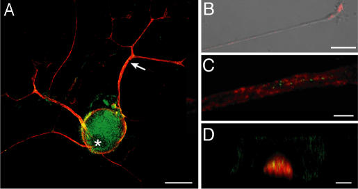
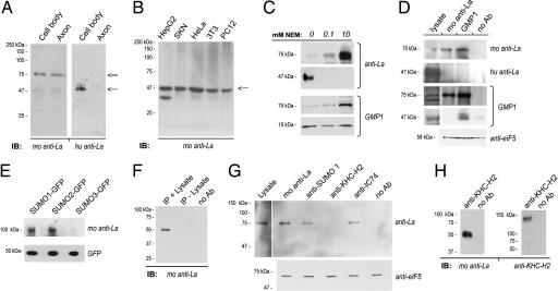
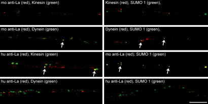
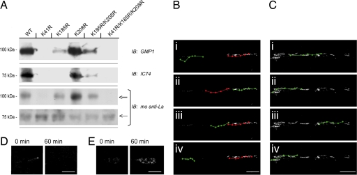
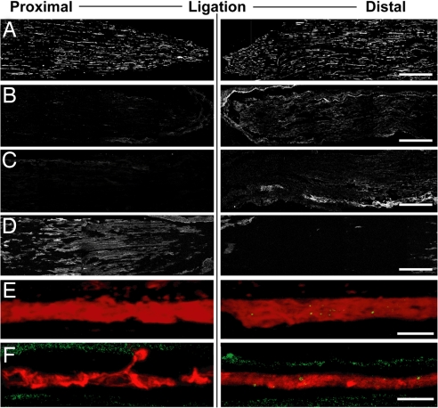
A surprisingly large population of mRNAs has been shown to localize to sensory axons, but few RNA-binding proteins have been detected in these axons. These axonal mRNAs include several potential binding targets for the La RNA chaperone protein. La is transported into axonal processes in both culture and peripheral nerve. Interestingly, La is posttranslationally modified in sensory neurons by sumoylation. In axons, small ubiquitin-like modifying polypeptides (SUMO)-La interacts with dynein, whereas native La interacts with kinesin. Lysine 41 is required for sumoylation, and sumoylation-incompetent La(K41R) shows only anterograde transport, whereas WT La shows both anterograde and retrograde transport in axons. Thus, sumoylation of La determines the directionality of its transport within the axonal compartment, with SUMO-La likely recycling to the cell body.


Conflict of interest statement

The authors declare no conflict of interest.

Figures

Fig. 1.
Fig. 1.
La autoantigen extends into regenerating sensory axons. (A) Cultures of injury-conditioned DRGs were colabeled with mouse anti-La (green) and chick antineurofilament (NF) antibodies (red). La is seen in the cell body, including the nucleus, and axonal processes (arrow). (B) In a single xy image of a distal axon, mouse anti-La (red) merged with DIC shows a granular La immunoreactivity. (C and D) Sections of sciatic nerve at ≈1 cm proximal to the crush site (7 d after injury) were colabeled for mouse anti-La (green) and chick anti-NF (red). C shows a single xy plane taken through the center of the axon. D shows a single xz plane demonstrating intraaxonal La with less-aggregated signals in the surrounding Schwann cell cytoplasm. (Scale bars: A, 40 μm; B, 20 μm; C and D, 5 μm.)
Fig. 2.
Fig. 2.
Neuronal La is sumoylated. (A) In immunoblots of cell body and axonal lysates from DRG cultures, immunoblotting with mouse anti-La shows a strong band migrating at ≈75 kDa (double arrow) and a faint band at ≈47 kDa (single arrow), whereas blotting with human anti-La only detects the lower band. (B) In blots prepared from HepG2, SKN, HeLa, NIH 3T3, and nondifferentiated PC12 cells lysed in standard RIPA buffer, mouse anti-La only detected a 47-kDa band (arrow). (C) Axoplasm from crushed sciatic nerve prepared in nondenaturing buffer supplemented NEM was used for immunoblotting. The mouse anti-La detected increasing amounts of 75-kDa La and decreasing amounts of 47-kDa La with increasing NEM concentration. The human anti-La showed a similar decrease in the 47-kDa La. Anti-SUMO1 (GMP1) showed increasing levels of a 75-kDa band with increasing NEM concentration. There was a slight decrease in the 17 kDa corresponding to free SUMO1 with increasing NEM concentration. (D) Axoplasm isolated in the presence of 10 mM NEM was used for immunoprecipitation with mouse and human anti-La or GMP1, as indicated. Mouse anti-La detects a 75-kDa band precipitated by GMP1. GMP1 recognized a sumoylated protein migrating at 75 kDa in the La precipitates, but nothing was detected at 47 kDa. The human anti-La did not recognize bands in the mouse anti-La or GMP1 precipitates. Supernatant from the precipitations probed with anti-eIF5 showed approximately equivalent levels of input protein for these samples. (E) Expression of SUMO-GFPs in DRG cultures followed by precipitation with rabbit anti-GFP shows an ≈100-kDa band for the SUMO1- and SUMO2-GFP-expressing cells when probed with mouse anti-La. The anti-La detects a faint band at ≈100 kDa in the SUMO3-GFP-expressing cells. Probing these blots with anti-GFP shows relatively equivalent expression of free SUMO-GFP. (F) Mouse anti-La immunoprecipitations were processed with or without lysate to test the specificity of the Trueblot reagent, which was used for G and H, for native vs. denatured mouse IgG. The latter would be present in both preparations, but antigen would be present only in the plus-lysate sample. Even with extended exposure, bands corresponding to La were detected only in the IP plus lysate sample. (G) DRG cultures were lysed in RIPA buffer supplemented with 10 mM NEM and used for immunoprecipitation with mouse anti-La, GMP1, anti-KHC-H2 (kinesin), and anti-IC74 (dynein) followed by immunoblotting for mouse anti-La. Seventy-five-kilodalton La-reactive bands (double arrow) are seen in the SUMO1 and dynein precipitates but not in the kinesin precipitates. Supernatant from the precipitations probed with anti-eIF5 showed approximately equivalent levels of input protein for these samples. (H) Axoplasm prepared in the absence of NEM and processed for precipitation with KHC2-H2 antibody. By immunoblotting, only 47-kDa La coprecipitates with kinesin heavy chain; reprobing with the anti-KHC2-H2 shows successful precipitation of kinesin heavy chain. Experiments performed in the presence of 10 mM NEM showed identical results (data not shown).
Fig. 3.
Fig. 3.
Regenerating axons contain SUMO that colocalizes with La and dynein. DRG cultures were colabeled with xenon-labeled GMP1, mouse anti-La, anti-KHC-H2, and anti-IC74, as indicated. Signals for human anti-La were detected by indirect immunofluorescence. Single xy planes of midaxon shaft of 24-h injury-conditioned DRG cultures are shown. Note that signals detected by the mouse anti-La show focal colocalization with dynein and SUMO1 (arrows). Conversely, the signals detected by the human anti-La, which does recognize SUMO-La, show focal colocalization with kinesin (arrows) but no overlap with SUMO1 or dynein. Signals for SUMO1 show focal colocalization with dynein (arrows) but no overlap with kinesin. (Scale bar: 10 μm.)
Fig. 4.
Fig. 4.
Lysine 41 is critical for sumoylation and retrograde transport of La. (A and B) Naive DRG cultures were transfected with WT and mutant huLa-GFP, as indicated. Anti-GFP immunoprecipitates were used for immunoblotting. Sumoylated and native huLa-GFP, detected by the mouse anti-La, could be discerned by distinct migration (double and single arrows, respectively). GMP1 blots showed a prominent band in the huLa-GFPWT and huLa-GFPK208R with fainter bands detected in the huLa-GFPK185R and huLa-GFPK185R/K208R. These samples also showed more abundant SUMO-huLaGFP (≈100 kDa) than native huLa-GFP (≈75 kDa) when probed with the anti-La. The huLa-GFPK41R and huLa-GFPK41R/K185R/K208R mutants showed no detectable signals for SUMO1 and showed more abundant native huLa-GFP than SUMO-huLa-GFP based on molecular mass. A similar pattern was seen for coprecipitating dynein detected with the IC74 antibody (i.e., huLa-GFPWT, huLa-GFPK208R > huLa-GFPK185R, huLa-GFPK185R/K208R > huLa-GFPK41R, and huLa-GFPK41R/K185R/K208R). (B and C) DRG neurons transfected with huLa-GFPWT (B) and huLa-GFPK41R (C) were used for live cell imaging. Images were collected over a 60-min period and i–iv display movement of individual GFP aggregates over this course (anterograde, green; retrograde, red). Note that huLa-GFPWT shows both anterograde and retrograde motility, but the huLa-GFPK41R mutant shows only ograde motility (also see SI Movies 1 and 2). (D and E) Exposure-matched images of growth cones sampled at time 0 and 60 min for huLa-GFPWT (D) and huLa-GFPK41R (E) are shown. Note that only huLa-GFPK41R mutant accumulates in the growth cone. (Scale bars: B and C, 10 μm; D and E, 5 μm.)
Fig. 5.
Fig. 5.
Sumoylated La accumulates distal to sciatic nerve ligation. Sections of ligated crushed sciatic nerve (3 d after injury and ligation) were processed for immunostaining and analyzed by confocal microscopy. Nerve segment proximal to the ligation is displayed on Left, and segment distal to the ligation is displayed on Right; crush site is Right. Montages of four free maximum projections are illustrated in each image of A–D (each projection was generated from 10 optical planes taken at 0.9 μm). E and F represent single optical planes through the central region of one representative axon from the corresponding regions. All image pairs are matched for laser power, photomultiplier tube voltage, offset, and processing. A displays the NF signal. Nerve sections stained with the mouse anti-La show a more prominent signal distal to the ligation site [B; also see merged NF (red) and anti-La (green) in E]. Staining with the GMP1 antibody showed a similar accumulation of SUMO1 distal to the ligation [C; also see merged NF (red) and GMP1 (green) in F]. Kinesin, detected by anti-KHC-H2 antibody, accumulated proximal to the lesion site (D). (Scale bars: A–D, 500 μm; E and F, 10 μm.)

References

    1. Twiss JL, van Minnen J. J Neurotrauma. 2006;23:295–308. - PubMed
    1. Hirokawa N. J Neurosci. 2006;26:7139–7142. - PMC - PubMed
    1. Wolin SL, Cedervall T. Annu Rev Biochem. 2002;71:375–403. - PubMed
    1. Maraia RJ, Bayfield MA. Mol Cell. 2006;21:149–152. - PubMed
    1. Fok V, Friend K, Steitz JA. J Cell Biol. 2006;173:319–325. - PMC - PubMed

Publication types

MeSH terms

LinkOut - more resources