. 2007 Jul 31;104(31):12790-5.
doi: 10.1073/pnas.0701437104. Epub 2007 Jul 23.

Elevated NF-kappaB responses and FLIP levels in leukemic but not normal lymphocytes: reduction by salicylate allows TNF-induced apoptosis

Affiliations

Elevated NF-kappaB responses and FLIP levels in leukemic but not normal lymphocytes: reduction by salicylate allows TNF-induced apoptosis

Colin Rae et al. Proc Natl Acad Sci U S A. .

Abstract

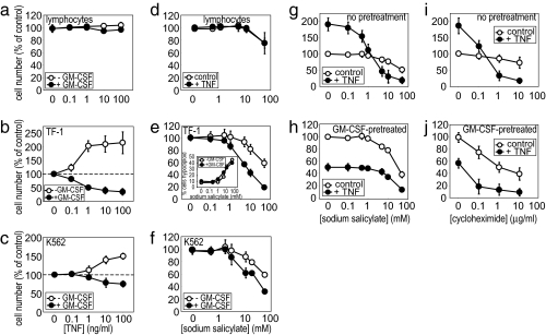
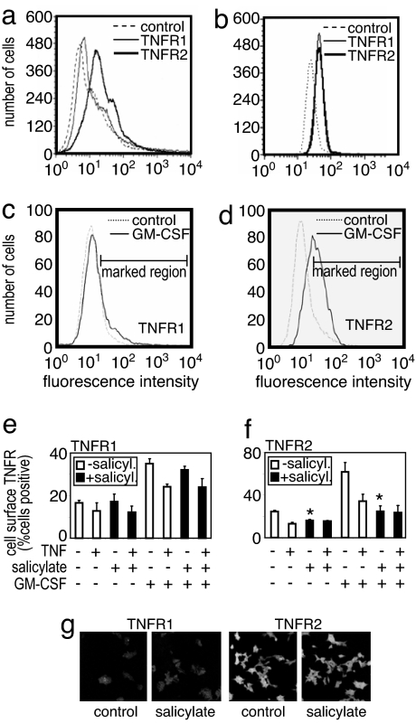
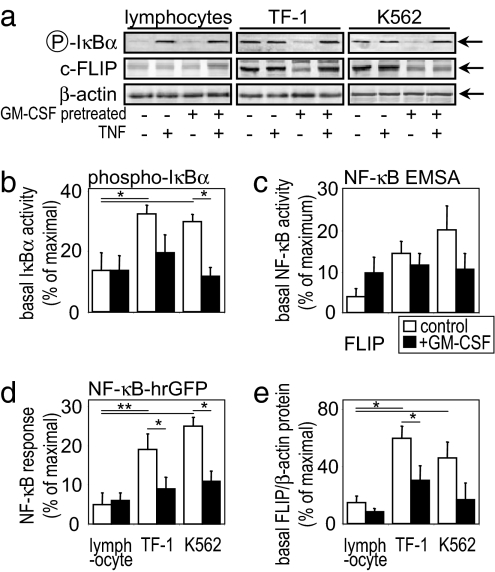
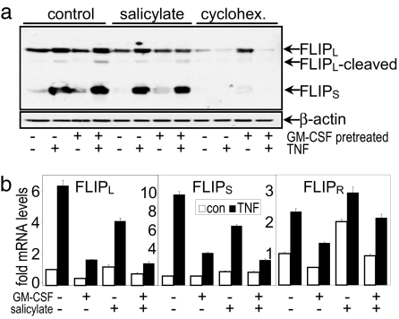
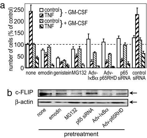
As its name suggests, tumor necrosis factor (TNF) is known to induce cytotoxicity in a wide variety of tumor cells and cell lines. However, its use as a chemotherapeutic drug has been limited by its deleterious side effects of systemic shock and widespread inflammatory responses. Some nonsteroidal antiinflammatory drugs, such as sodium salicylate, have been shown to have a chemopreventive role in certain forms of cancer. Here, we reveal that sodium salicylate selectively enhances the apoptotic effects of TNF in human erythroleukemia cells but does not affect primary human lymphocytes or monocytes. Sodium salicylate did not affect the intracellular distribution of TNF receptors (TNFRs) but stimulated cell surface TNFR2 shedding. Erythroleukemia cells were shown to possess markedly greater basal NF-kappaB responses and elevated Fas-associated protein with death domain-like IL-1 converting enzyme (FLIP) levels. Sodium salicylate achieved its effects by reducing the elevated NF-kappaB responsiveness and FLIP levels and restoring the apoptotic response of TNF rather than the proliferative/proinflammatory effects of the cytokine in these cancer cells. Inhibition of NF-kappaB or FLIP levels in human erythroleukemia cells by pharmacological or molecular-biological means also resulted in switching the character of these cells from a TNF-responsive proliferative phenotype into an apoptotic one. These findings expose that the enhanced proliferative nature of human leukemia cells is caused by elevated NF-kappaB and FLIP responses and basal levels, reversible by sodium salicylate to allow greater apoptotic responsiveness of cytotoxic stimuli such as TNF. Such findings provide insight into the molecular mechanisms by which human leukemia cells can switch from a proliferative into an apoptotic phenotype.


Conflict of interest statement

The authors declare no conflict of interest.

Figures

Fig. 1.
Fig. 1.
TNF, sodium salicylate, and cycloheximide responses in normal and leukemic cells. MTS [3-(4,5-dimethylthiazol-2-yl)-5-(3-carboxymethoxyphenyl)-2-(4-sulfophenyl)-2H-tetrazolium] proliferation/cytotoxicity assays were performed on primary lymphocytes (a and d) and TF-1 (b, e, g–j), and K562 (c and f) leukemia cells treated with TNF (a–c), sodium salicylate (d–f, i, and j) or cycloheximide (g and h) stimulation. Except where indicated, cells were pretreated with 1 ng/ml GM-CSF or 50 ng/ml TNF for 24 h before stimulation. Data represent the means ± SEM, n = 4 (n = 3 for i and j).
Fig. 2.
Fig. 2.
Sodium salicylate alteration of TNFR expression. Primary human lymphocytes (a) and TF-1 cells (b–f) treated as indicated, had their cell surface (a–f) or intracellular (g) TNFR1 and TNFR2 levels measured. TNFR levels of basal (a and b), 24 h, 1 ng/ml GM-CSF-pretreated (c–f), or sodium salicylate-treated (e–g) cells showed that GM-CSF increased TNFR2 expression that could be shed by TNF (50 ng/ml, 30 min) or sodium salicylate (5 mM, 1 h) stimuli. Primary human lymphocytes showed no sodium salicylate-induced shedding of TNFRs (data not shown). (g) TNFR subcellular distribution in HeLa-TNFR2 cells, with TF-1 cells similarly showing no subcellular alteration by sodium salicylate (data not shown). The data are from a single representative experiment repeated at least three other times with similar findings. Histograms represent the means ± SEM., n = 4, of immunofluorescence intensities within the marked regions (c and d).
Fig. 3.
Fig. 3.
Elevated NF-κB responsiveness in leukemia cells. (a) Western blot analysis of primary lymphocytes and TF-1 and K562 cells pretreated for 24 h with 1 ng/ml GM-CSF and/or 50 ng/ml TNF treatment for a further 24 h where indicated. Quantification of cellular IκBα protein levels (b), p65 NF-κB DNA-binding ELISA (c), NF-κB transcription reporter construct activity (d), and c-FLIP levels (e) is shown. Total β-actin protein levels were used as a loading control. Data represent the means ± SEM of at least four separate experiments.
Fig. 4.
Fig. 4.
Sodium salicylate reduces elevated FLIP levels in leukemia cells. (a) Western blot analysis of TF-1 cells pretreated for 24 h with 1 ng/ml GM-CSF and/or 50 ng/ml TNF for a further 4 h where indicated. Where shown, cells had 5 mM sodium salicylate or 1 μg/ml cycloheximide preincubated for 1 h or 24 h, respectively, before the addition of TNF stimulus. FLIP isoforms were detected by NF-6 antibody. Concurrently run positive controls confirmed the identity of the bands, which are indicated by arrows. (b) Quantification of cellular FLIPL, FLIPS, and FLIPR mRNA levels by real-time PCR. Data represent the means ± SEM of at least six separate experiments.
Fig. 5.
Fig. 5.
Inhibition of NF-κB and FLIP levels in leukemia cells. (a) MTS assay of TF-1 cells pretreated with the indicated pharmacological inhibitor (30 μg/ml emodin, 25 μg/ml genistein, or 5 μM MG132) for 1 h before 50 ng/ml TNF treatment for a further 24 h. Where indicated, cells were pretreated for 24 h with 1 ng/ml GM-CSF. p65 NF-κB siRNA and adenoviral pretreatments were for 24 h before TNF stimulation. Data represent the means ± SEM of at least three separate experiments. (b) c-FLIP and β-actin protein levels in pretreated TF-1 cells. The Western blotting data are from a single experiment representative of at least three independent results.

References

    1. Carswell EA, Old LJ, Kassel RL, Green S, Fiore N, Williamson B. Proc Natl Acad Sci USA. 1975;72:3666–3670. - PMC - PubMed
    1. Fiers W. FEBS Lett. 1991;285:199–212. - PubMed
    1. Berkova N, Lemay A, Korobko V, Shingarova L, Sagaidak L, Goupil S. Cancer Detection Prevention. 1999;23:1–7. - PubMed
    1. MacEwan DJ. Cell Signalling. 2002;14:477–492. - PubMed
    1. Dannenberg AJ, Altorki NK, Boyle JO, Dang C, Howe LR, Weksler BB, Subbararnaiah K. Lancet Oncol. 2001;2:544–551. - PubMed

Publication types

MeSH terms

Substances