

Adoptive transfer of autologous, HER2-specific, cytotoxic T lymphocytes for the treatment of HER2-overexpressing breast cancer
- PMID: 17646988
- PMCID: PMC11030865
- DOI: 10.1007/s00262-007-0355-7
Adoptive transfer of autologous, HER2-specific, cytotoxic T lymphocytes for the treatment of HER2-overexpressing breast cancer
Abstract
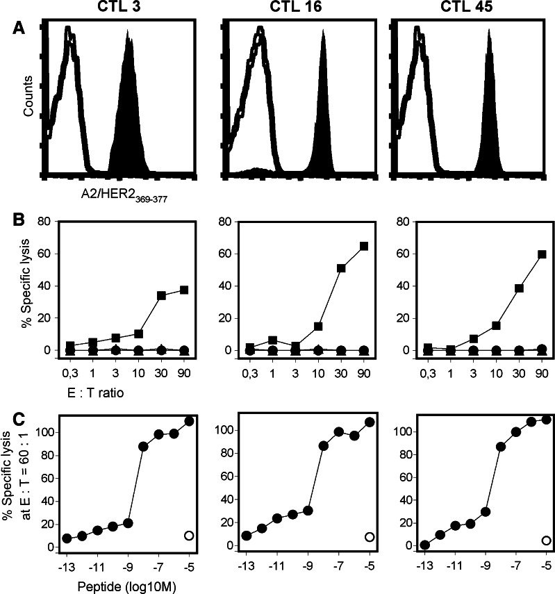
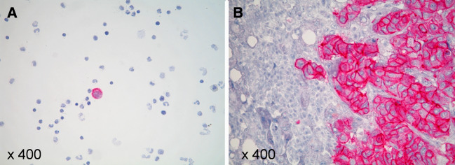
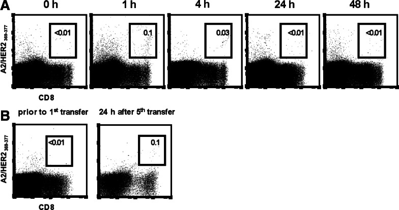
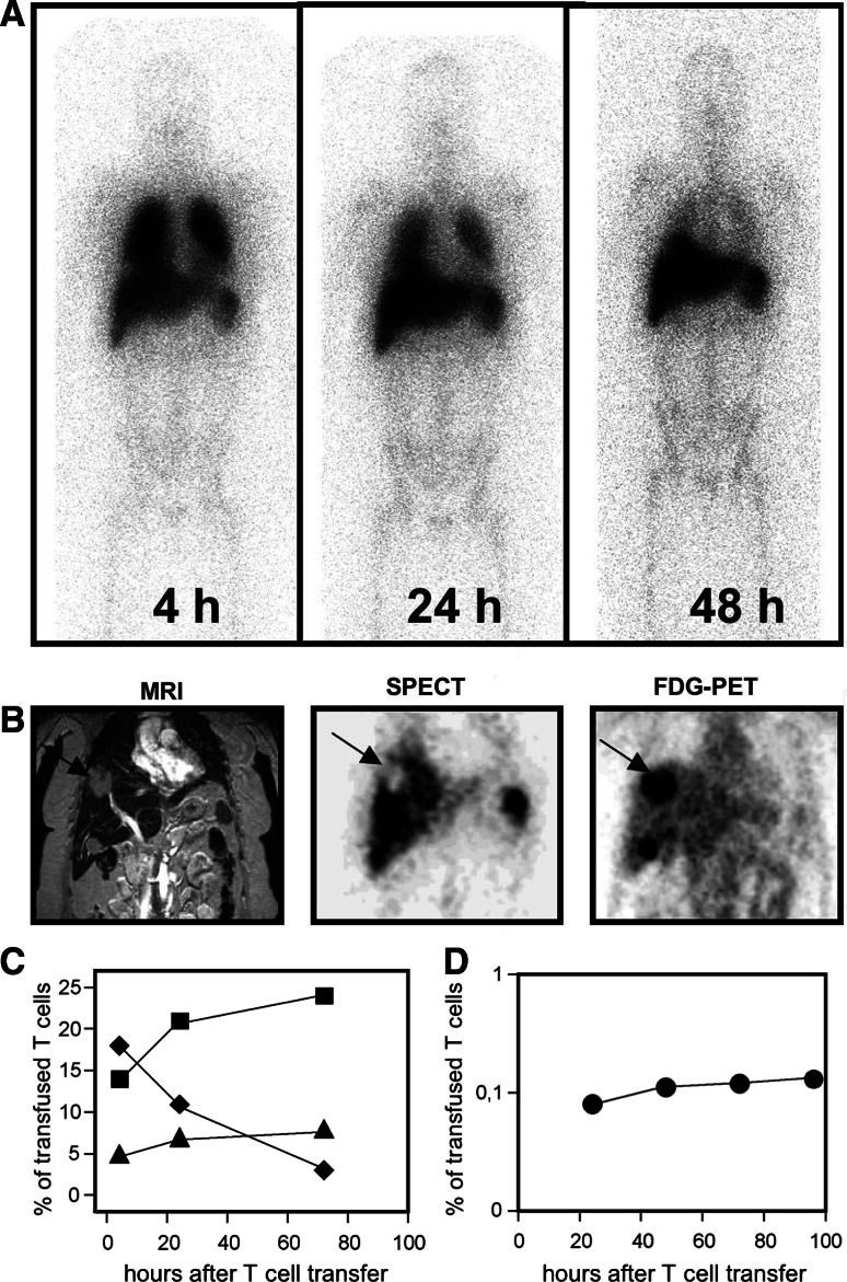
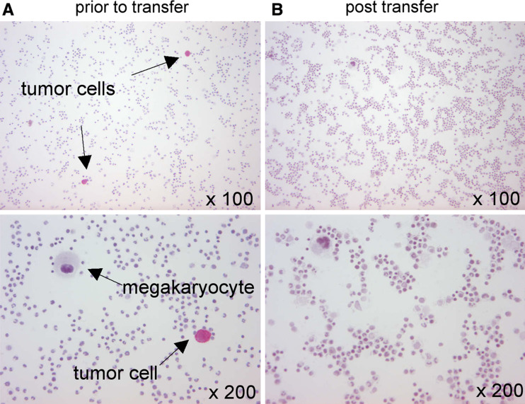
The human epidermal growth factor receptor 2 (HER2) has been targeted as a breast cancer-associated antigen by immunotherapeutical approaches based on HER2-directed monoclonal antibodies and cancer vaccines. We describe the adoptive transfer of autologous HER2-specific T-lymphocyte clones to a patient with metastatic HER2-overexpressing breast cancer. The HLA/multimer-based monitoring of the transferred T lymphocytes revealed that the T cells rapidly disappeared from the peripheral blood. The imaging studies indicated that the T cells accumulated in the bone marrow (BM) and migrated to the liver, but were unable to penetrate into the solid metastases. The disseminated tumor cells in the BM disappeared after the completion of adoptive T-cell therapy. This study suggests the therapeutic potential for HER2-specific T cells for eliminating disseminated HER2-positive tumor cells and proposes the combination of T cell-based therapies with strategies targeting the tumor stroma to improve T-cell infiltration into solid tumors.
Figures
Comment in
-
Tumor stromal barriers to the success of adoptive T cell therapy.Cancer Immunol Immunother. 2008 Feb;57(2):281-3. doi: 10.1007/s00262-007-0356-6. Epub 2007 Jul 24. Cancer Immunol Immunother. 2008. PMID: 17646987 Free PMC article. No abstract available.
Similar articles
-
Generation of populations of antigen-specific cytotoxic T cells using DCs transfected with DNA construct encoding HER2/neu tumor antigen epitopes.BMC Immunol. 2017 Jun 20;18(1):31. doi: 10.1186/s12865-017-0219-7. BMC Immunol. 2017. PMID: 28633645 Free PMC article.
-
The generation of both T killer and Th cell clones specific for the tumor-associated antigen HER2 using retrovirally transduced dendritic cells.J Immunol. 2001 Aug 1;167(3):1712-9. doi: 10.4049/jimmunol.167.3.1712. J Immunol. 2001. PMID: 11466395
-
Antihuman epidermal growth factor receptor 2 (HER2) monoclonal antibody trastuzumab enhances cytolytic activity of class I-restricted HER2-specific T lymphocytes against HER2-overexpressing tumor cells.Cancer Res. 2002 Apr 15;62(8):2244-7. Cancer Res. 2002. PMID: 11956077
-
Adoptive immuno-gene therapy of cancer with single chain antibody [scFv(Ig)] gene modified T lymphocytes.J Biol Regul Homeost Agents. 2004 Apr-Jun;18(2):134-40. J Biol Regul Homeost Agents. 2004. PMID: 15471217 Review.
-
Is antigen specificity the key to efficient adoptive T-cell therapy?Immunotherapy. 2011 Apr;3(4):495-505. doi: 10.2217/imt.11.16. Immunotherapy. 2011. PMID: 21463191 Review.
Cited by
-
Surmounting the obstacles that impede effective CAR T cell trafficking to solid tumors.J Leukoc Biol. 2020 Oct;108(4):1067-1079. doi: 10.1002/JLB.1MR0520-746R. Epub 2020 Jul 3. J Leukoc Biol. 2020. PMID: 32620049 Free PMC article. Review.
-
Glioblastoma, an opportunity T cell trafficking could bring for the treatment.Mol Biol Rep. 2022 Oct;49(10):9863-9875. doi: 10.1007/s11033-022-07510-1. Epub 2022 May 23. Mol Biol Rep. 2022. PMID: 35604627 Review.
-
A transductionally retargeted adenoviral vector for virotherapy of Her2/neu-expressing prostate cancer.Hum Gene Ther. 2012 Jan;23(1):70-82. doi: 10.1089/hum.2011.016. Epub 2011 Oct 12. Hum Gene Ther. 2012. PMID: 21875358 Free PMC article.
-
Tumor Microenvironment Immunosuppression: A Roadblock to CAR T-Cell Advancement in Solid Tumors.Cells. 2022 Nov 16;11(22):3626. doi: 10.3390/cells11223626. Cells. 2022. PMID: 36429054 Free PMC article. Review.
-
Analysis of scRNA-seq and bulk RNA-seq demonstrates the effects of EVI2B or CD361 on CD8+ T cells in osteosarcoma.Exp Biol Med (Maywood). 2023 Jan;248(2):130-145. doi: 10.1177/15353702221142607. Epub 2022 Dec 13. Exp Biol Med (Maywood). 2023. PMID: 36511103 Free PMC article.
References
-
- Jäger E, Nagata Y, Gnjatic S, Wada H, Stockert E, Karbach J, Dunbar PR, Lee SY, Jungblut A, Jäger D, Arand M, Ritter G, Cerundolo V, Dupont B, Chen Y-T, Old LJ, Knuth A. Monitoring CD8 T cell responses to NY-ESO-1: correlation of humoral and cellular immune responses. Proc Natl Acad Sci USA. 2000;97:4760–4765. doi: 10.1073/pnas.97.9.4760. - DOI - PMC - PubMed
Publication types
MeSH terms
LinkOut - more resources
Full Text Sources
Other Literature Sources
Medical
Research Materials
Miscellaneous
