. 2007 Nov 1;407(3):407-17.
doi: 10.1042/BJ20070195.

Regulation of stress-induced intracellular sorting and chaperone function of Hsp27 (HspB1) in mammalian cells

Affiliations

Regulation of stress-induced intracellular sorting and chaperone function of Hsp27 (HspB1) in mammalian cells

Anton L Bryantsev et al. Biochem J. .

Abstract

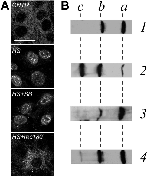
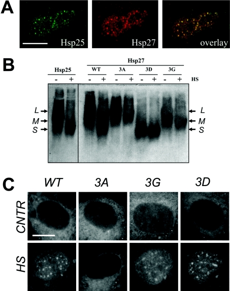
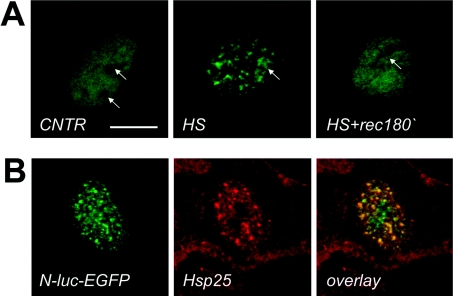
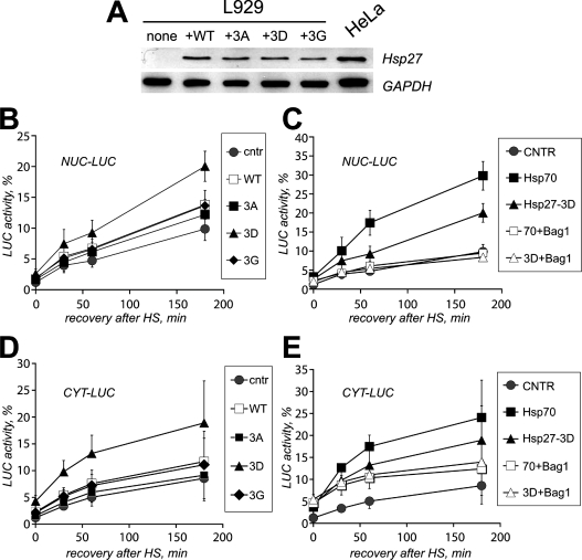
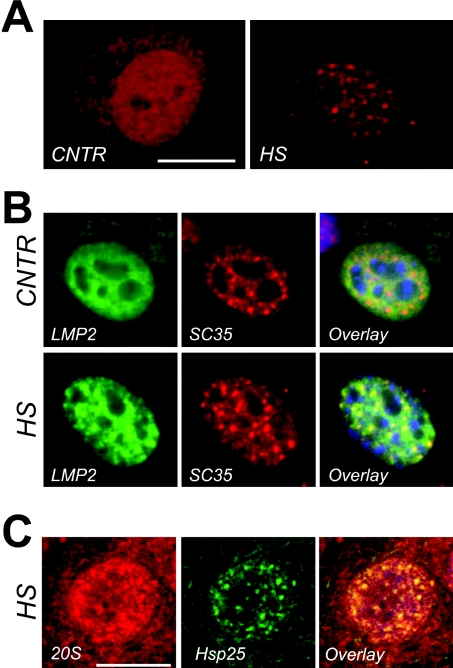
In vitro, small Hsps (heat-shock proteins) have been shown to have chaperone function capable of keeping unfolded proteins in a form competent for Hsp70-dependent refolding. However, this has never been confirmed in living mammalian cells. In the present study, we show that Hsp27 (HspB1) translocates into the nucleus upon heat shock, where it forms granules that co-localize with IGCs (interchromatin granule clusters). Although heat-induced changes in the oligomerization status of Hsp27 correlate with its phosphorylation and nuclear translocation, Hsp27 phosphorylation alone is not sufficient for effective nuclear translocation of HspB1. Using firefly luciferase as a heat-sensitive reporter protein, we demonstrate that HspB1 expression in HspB1-deficient fibroblasts enhances protein refolding after heat shock. The positive effect of HspB1 on refolding is completely diminished by overexpression of Bag-1 (Bcl-2-associated athanogene), the negative regulator of Hsp70, consistent with the idea of HspB1 being the substrate holder for Hsp70. Although HspB1 and luciferase both accumulate in nuclear granules after heat shock, our results suggest that this is not related to the refolding activity of HspB1. Rather, granular accumulation may reflect a situation of failed refolding where the substrate is stored for subsequent degradation. Consistently, we found 20S proteasomes concentrated in nuclear granules of HspB1 after heat shock. We conclude that HspB1 contributes to an increased chaperone capacity of cells by binding unfolded proteins that are hereby kept competent for refolding by Hsp70 or that are sorted to nuclear granules if such refolding fails.


Figures

Figure 1
Figure 1. Distribution and phosphorylation patterns of endogenous HspB1 (Hsp25) in Rat-1 fibroblasts before, during and after heat shock
Localization of Hsp25 in cells (A) or phosphorylation status of Hsp25 in cell lysates (B) at normal conditions (CNTR, lane 1), after heat shock at 43 °C for 30 min without (HS, lane 2) or with (HS+SB, lane 3) the kinase inhibitor SB202190, or after heat shock followed by a recovery for 180 min at 37 °C (HS+rec180′, lane 4). (A) Confocal images of cells immunostained with the antibody against rodent Hsp25. Bar=30 μm. (B) Cells were lysed and run on 7% acrylamide IEF gels and processed for Western blot analysis using the antibody against rodent Hsp25. Positions of non-phosphorylated (a), monophosphorylated (b), and diphosphorylated (c) isoforms are indicated.
Figure 2
Figure 2. Ultrastructural immunolocalization of Hsp25 in the nuclei of heat-shocked Rat-1 fibroblasts
To identify the intranuclear localization of rodent Hsp25 after heating at the ultrastructural level, heated (30 min at 43 °C) Rat-1 cells were briefly lysed in TEA buffer containing Mg2+ prior to standard fixation in paraformaldehyde. After immunolabelling with a primary anti-Hsp25 antibody and a secondary goat anti-rabbit 5 nm gold-labelled antibody, ultrathin sections of the cells were processed for transmission electron microscopy. (A) Entire cell nucleus of a cell heat shocked for 30 min. Arrows indicate IGCs and the arrowhead indicates the nucleolus. The inset represents a typical nucleus of a similarly treated cell, stained for immunofluorescence. Bar=1 μm. (B) Enlarged image of an IGC clearly enriched with the Hsp27 label. Bar=1 μm. (C) Enlarged fragment of a perichromatin fibre, containing the label along its periphery. Bar=1 μm.
Figure 3
Figure 3. Intracellular distribution and oligomeric states of endogenous Hsp25 and ectopically expressed WT and mutants of human Hsp27 in Rat-1 fibroblasts
(A) Rat-1 cells were transiently transfected with a plasmid encoding human Hsp27, heat shocked and processed for immunofluorescence analysis using confocal microscopy. Endogenous rat Hsp25 (green) and ectopically expressed human Hsp27 (red) and the merged image show their excellent co-localization (overlay: yellow). Bar=10 μm. (B) Rat-1 cells were transiently transfected with plasmids encoding WT human Hsp27 or its mutants Hsp27-3A (3A), Hsp27-3G (3G), and Hsp27-3D (3D). The oligomeric size distribution of the endogenous rodent Hsp25 and ectopically expressed Hsp27 variants was analysed in cells before (−) and after (+) heat shock at 43 °C for 30 min (HS). Cell lysates were run by native non-reducing pore size-exclusion electrophoresis, blotted on to nitrocellulose membrane and probed with species-specific anti-Hsp antibodies, recognizing either Hsp25 or Hsp27. Arrows show three major intermediates in oligomers corresponding to their large (L), medium (M) and small (S) size. For details on the various Hsp27 phosphorylation mutants see the text. (C) Rat-1 cells were transiently transfected with plasmids encoding WT human Hsp27 or its mutants Hsp27-3A (3A), Hsp27-3G (3G) and Hsp27-3D (3D) and their intracellular localization was analysed using confocal microscopy as described in (A), before (CNTR) and after heat shock (HS). Bar=10 μm.
Figure 4
Figure 4. Localization of the nuclear stress-sensitive luciferase reporter before and after heating Rat-1 cells
(A) Rat-1 cells were transiently transfected with plasmids encoding EGFP-tagged nuclear luciferase (N-luc-EGFP) and either left unheated (CNTR), or heated for 30 min at 43 °C (HS) or heated and allowed to recover for 180 min at 37 °C (HS+rec180′) before processing to confocal microscopy using EGFP as a tracer. Arrows indicate the position of the nucleoli. Note the similarity in kinetic behaviour after heat shock between nuclear luciferase and Hsp25 (Figure 1). Bar=10 μm. (B) Rat-1 cells transiently expressing N-luc-EGFP were heat shocked before processing to confocal microscopy using EGFP as a tracer for luciferase localization (green) or anti-Hsp25 immunostaining (red). The merged image clearly demonstrates an excellent co-localization (overlay: yellow). DNA was counterstained with DAPI in blue.
Figure 5
Figure 5. Hsp27 overexpression enhances refolding of heat-inactivated luciferase reporter in L929 cells
L929 cells lacking endogenous Hsp25 were transiently transfected with plasmids encoding EGFP-tagged nuclear luciferase (AC) or cytosolic luciferase (D, E) and co-transfected with an empty plasmid (CNTR) or with plasmids encoding WT human Hsp27 or its mutants Hsp27-3A (3A), Hsp27-3G (3G), or Hsp27-3D (3D). (A) Expression levels of WT Hsp27 and its mutants in L929 upon transient transfection as detected by Western blotting (upper lanes). Transfection efficiencies were equal for all plasmids used. Extracts of HeLa cells, naturally expressing moderate levels of human Hsp27, are provided as a positive control. Immunostaining for GAPDH (glyceraldehyde-3-phosphate dehydrogenase) was used to demonstrate equal loading between lanes (bottom panel). (B) L929 cells expressing nuclear luciferase were heated (30 min at 43 °C) and cells were lysed 0–180 min after heating. CHX (cycloheximide, 20 μg/ml) was added just prior to heating to prevent new luciferase synthesis. Recovery of luciferase activity is indicated for L929 cells co-transfected with an empty vector (●) or co-transfected with WT human Hsp27 (□), Hsp27-3A (■), Hsp27-3G (♦), and Hsp27-3D (▲). Activity was calculated as a percentage of the initial luciferase activity before heat shock. (C) Same as (B), but here recovery of nuclear luciferase activity is depicted for cells co-transfected with an empty vector (●), Hsp27-3D alone (▲), Hsp27-3D and Bag-1 (Δ), Hsp70 alone (■) and Hsp70 and Bag-1 (□). (D) Same as (B), now showing recovery of cytosolic luciferase activity (CYT-LUC). (E) Same as (C), now showing recovery of cytosolic luciferase activity (CYT-LUC).
Figure 6
Figure 6. Association between nuclear granules of Hsp25, nuclear speckles and the 20S proteasomal machine
(A) Confocal images of cell nuclei decorated with anti-20S proteasomal antibodies in the nuclei of cells before (CNTR) and 60 min after a 30 min heat shock at 43 °C (HS). Bar=10 μm. (B) Multicolour channel images of nuclei, expressing GFP-labelled 20S core subunit LMP2 before (panel labelled CNTR) and after a 30-min heat shock at 43 °C (panel labelled HS). Each horizontal series represent the same nucleus in which epigenetical LMP2–GFP (LMP: green) and endogenous SC35 (SC35: red) are revealed and overlaid (Overlay: co-localization is evident by the presence of yellow colour). 20S proteasomes (LMP) become concentrated in nuclear speckles (SC35) after heat shock. Bar=5 μm. (C) Confocal images of heat-shock cells immunostained with the anti-Hsp25 antibody (Hsp25: green) and the anti-20S proteasome antibody (20S: red). Co-localization is evident in the overlay image (Overlay: yellow). On the overlaid images nuclear outline is indicated with blue (DAPI staining). Bar=10 μm.

Similar articles

Cited by

References

    1. Landry J., Chretien P., Lambert H., Hickey E., Weber L. A. Heat shock resistance conferred by expression of the human HSP27 gene in rodent cells. J. Cell Biol. 1989;109:7–15. - PMC - PubMed
    1. Lavoie J. N., Gingras-Breton G., Tanguay R. M., Landry J. Induction of Chinese hamster HSP27 gene expression in mouse cells confers resistance to heat shock. HSP27 stabilization of the microfilament organization. J. Biol. Chem. 1993;268:3420–3429. - PubMed
    1. Bryantsev A. L., Loktionova S. A., Ilyinskaya O. P., Tararak E. M., Kampinga H. H., Kabakov A. E. Distribution, phosphorylation, and activities of Hsp25 in heat-stressed H9c2 myoblasts: a functional link to cytoprotection. Cell Stress. Chaperones. 2002;7:146–155. - PMC - PubMed
    1. Jakob U., Gaestel M., Engel K., Buchner J. Small heat shock proteins are molecular chaperones. J. Biol. Chem. 1993;268:1517–1520. - PubMed
    1. Ehrnsperger M., Buchner J., Gaestel M. Structure and function of small heat-shock proteins. In: Fink A., Goto Y., editors. Molecular chaperones in the life cycle of proteins. New York: Marcel Dekker, Inc.; 1997. pp. 533–575.

Publication types