. 2007 Oct 15;407(2):171-7.
doi: 10.1042/BJ20070552.

Activation-coupled membrane-type 1 matrix metalloproteinase membrane trafficking

Affiliations

Activation-coupled membrane-type 1 matrix metalloproteinase membrane trafficking

Yi I Wu et al. Biochem J. .

Abstract

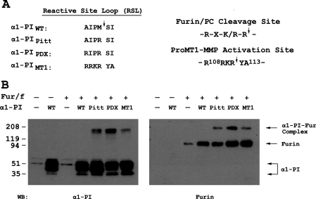
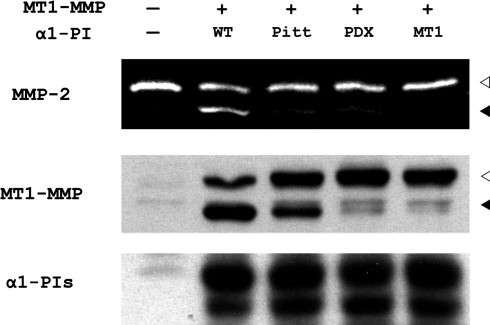
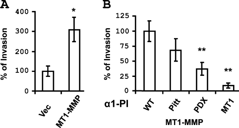
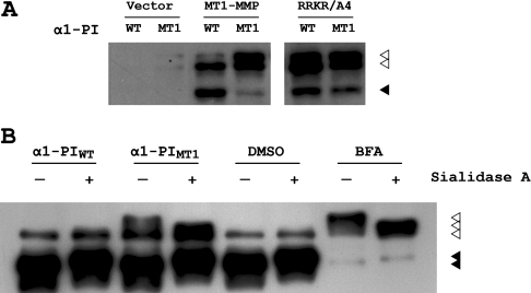
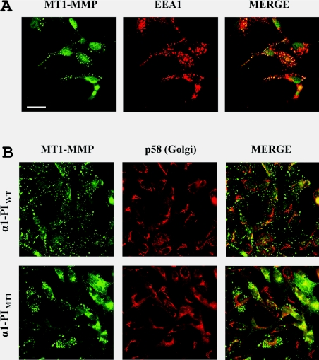
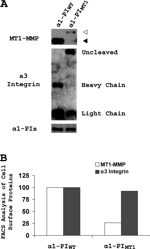
The transmembrane collagenase MT1-MMP (membrane-type 1 matrix metalloproteinase), also known as MMP-14, has a critical function both in normal development and in cancer progression, and is subject to extensive controls at the post-translational level which affect proteinase activity. As zymogen activation is crucial for MT1-MMP activity, an alpha1-PI (alpha1-proteinase inhibitor)-based inhibitor was designed by incorporating the MT1-MMP propeptide cleavage sequence into the alpha1-PI reactive-site loop (designated alpha1-PI(MT1)) and this was compared with wild-type alpha1-PI (alpha1-PI(WT)) and the furin inhibitory mutant alpha1-PI(PDX). Alpha1-PI(MT1) formed an SDS-stable complex with furin and inhibited proMT1-MMP activation. A consequence of the loss of MT1-MMP activity was the activation of proMMP-2 and the inhibition of MT1-MMP-mediated collagen invasion. alpha1-PI(MT1) expression also resulted in the intracellular accumulation of a glycosylated species of proMT1-MMP that was retained in the perinuclear region, leading to significantly decreased cell-surface accumulation of proMT1-MMP. These observations suggest that both the subcellular localization and the activity of MT1-MMP are regulated in a coordinated fashion, such that proMT1-MMP is retained intracellularly until activation of its zymogen, then proMT1-MMP traffics to the cell surface in order to cleave extracellular substrates.


Figures

Figure 1
Figure 1. A novel α1-PI variant based on the amino acid sequence flanking the proMT1-MMP activation site forms a SDS-stable complex with furin
(A) The amino acid sequence flanking the proMT1-MMP-activation site contains the optimal cleavage motif for furin and PC proteinases. A novel α1-PI variant, designated α1-PIMT1, was designed based on the sequence (P4–P2′) flanking the proMT1-MMP activation site. The altered sequence in the reactive-site loop (RSL) of α1-PIMT1 (P4–P2′) was aligned with the corresponding sequence of α1-PIWT and those of two other known α1-PI variants, α1-PIPitt and α1-PIPDX. (B) COS-7 cells were transiently transfected with a DNA plasmid encoding FLAG-tagged furin (Fur/f) in combination with plasmids encoding α1-PIWT (WT) or one of its variants α1-PIPitt (Pitt), α1-PIPDX (PDX) and α1-PIMT1 (MT1) respectively. Empty vectors (−) were used as controls in transfection. The cell lysates were prepared 24 h after transfection as described in the Experimental section and were fractionated by SDS/PAGE (5–20% gels) in duplicate, followed by Western blot analysis using an anti-α1-PI antibody (left-hand side) and anti-FLAG antibody (right-hand side). The immunopreciptates corresponding to α1-PI and furin alone and the α1-PI–furin (α1-PI–Fur) complex are indicated with arrows on the right-hand side.
Figure 2
Figure 2. α1-PIMT1 inhibits proMT1-MMP activation and prevents MT1-MMP-catalysed proMMP-2 activation
COS-7 cells were transiently transfected in duplicate with a DNA plasmid encoding FLAG-tagged MT1-MMP in combination with plasmids encoding α1-PIWT (WT) or different α1-PI variants α1-PIPitt (Pitt), α1-PIPDX (PDX) and α1-PIMT1 (MT1) respectively. The control cells (lane 1) were transfected with empty vectors (−). One set of transfected cells was incubated with 1 nM purified proMMP-2 in serum-free medium for 6 h, 18 h after transfection. The conditioned medium was analysed by gelatin zymography (top panel) as described in the Experimental section. The gelatinase activities corresponding to active MMP-2 and proMMP-2 are indicated with black and white arrowheads (top panel) respectively. The second set of transfected cells were analysed by SDS/PAGE, followed by Western blot analysis using an anti-FLAG antibody to detect MT1-MMP (middle panel) and an anti-(α1-PI) antibody (bottom panel) respectively. The immunoprecipitates corresponding to active MT1-MMP and proMT1-MMP are indicated with black and white arrowheads respectively.
Figure 3
Figure 3. α1-PIMT1 inhibits MT1-MMP-mediated collagen invasion
COS-7 cells were transiently transfected with (A) a vector or plasmid encoding MT1-MMP, or (B) with a plasmid encoding MT1-MMP in combination with a second plasmid encoding α1-PIWT or α1-PIPitt (Pitt), α1-PIPDX (PDX) and α1-PIMT1 (MT1) as indicated (right-hand side). After 24 h, the cells were analysed for three-dimensional collagen gel invasion as described in the Experimental section. Gel invasion is expressed as percentage invasion relative to cells transfected with vector (A) or cells transfected with both MT1-MMP and α1-PIWT (B).
Figure 4
Figure 4. α1-PIMT1 inhibition of proMT1-MMP activation results in the accumulation of glycosylated proMT1-MMP
(A) COS-7 cells were transiently transfected with a DNA plasmid encoding FLAG-tagged MT1-MMP or proMT1-MMP containing the activation site mutated to alanine residues (RRKR/A4), in combination with α1-PIWT or α1-PIMT1 respectively. Empty vector was used as a control. After 24 h, the cells were lysed, resolved by SDS/PAGE (10% gels) and analysed by Western blotting using an anti-FLAG antibody. The immunoprecipitates corresponding to activated MT1-MMP and proMT1-MMP are indicated with black and white arrowheads respectively. (B) COS-7 cells were transiently transfected with a DNA plasmid encoding FLAG-tagged MT1-MMP and also transfected with either α1-PIWT or α1-PIMT1, or were incubated with 10 μg/ml BFA (with DMSO used as vehicle control) respectively. The FLAG-tagged proteins were purified using anti-FLAG M2–agarose, followed by in vitro enzymatic de-glycosylation with sialydase A. The reaction mixtures were fractionated by SDS/PAGE (10% gels), followed by Western blot analysis using a polyclonal anti-[MT1-MMP (hinge region)] antibody. The immunoprecipitates corresponding to activated MT1-MMP and proMT1-MMP are indicated with black and white arrowheads respectively.
Figure 5
Figure 5. α1-PIMT1 induces perinuclear accumulation of endogenous MT1-MMP in MDA-MB-231 cells
(A) Cells were transfected with α1-PIWT and after 24 h, the cells were fixed with 3.7% (v/v) formalin and permeablized using 0.1% (v/v) Triton X-100. and immunostained using a rabbit anti-[MT1-MMP (hinge region)] antibody and a monoclonal mouse anti-EEA1 (early endosome antigen 1) antibody (B) Cells were stably transfected with α1-PIWT or α1-PIMT1 and, after 24 h, the cells were fixed with 3.7% (v/v) formalin and permeablized using 0.1% (v/v) Triton X-100. The cells were immunostained using a rabbit anti-[MT1-MMP (hinge region)] antibody and a monoclonal mouse anti-p58 (Golgi marker) monoclonal antibody. In both (A) and (B), the primary immunostaining was followed by Alexa Fluor® 488 -conjugated goat anti-(rabbit IgG) and Alexa Fluor® 546-conjugated goat anti-(mouse IgG) secondary antibodies. The images were acquired using a Zeiss LSM510 laser-scanning confocal microscope. Overlay images (MERGE) were generated using Adobe Photoshop software. Scale bar, 20 μm.
Figure 6
Figure 6. α1-PIMT1 inhibits cell-surface presentation of MT1-MMP
(A) MDA-MB-231 cells were stably transfected with either α1-PIWT or α1-PIMT1. The cell-surface proteins were biotinylated and purified as described in the Experimental section. The purified cell-surface proteins were fractionated in duplicate by SDS/PAGE (5–20% gels) under reducing conditions, followed by Western blot analysis using an anti-[MT1-MMP (hinge region)] antibody (top) and a mixture of anti-(α3 integrin heavy chain) and anti-(α3 integrin light chain) antibodies (middle) respectively. The cell lysate inputs before purification were analysed by Western blotting using an anti-α1-PI antibody (bottom). The immunoprecipitates corresponding to activated MT1-MMP and proMT1-MMP are indicated with black and white arrowheads respectively. (B) MDA-MB-231 cells stably expressing α1-PIWT or α1-PIMT1 were labelled on ice with an anti-[MT1-MMP (hinge region)] antibody and an anti-(α3 integrin) antibody (ASC-6), followed by staining with Alexa Fluor® 488- and 546-conjugated secondary antibodies respectively. The labelled cells were analysed by FACS for fluorescence intensities on both wavelengths. The means of fluorescence intensities for MT1-MMP and α3 integrin in the α1-PIMT1 cells are are compared with α1-PIWT cells (100%).

References

    1. Sternlicht M. D., Werb Z. How matrix metalloproteinases regulate cell behavior. Annu. Rev. Cell Dev. Biol. 2001;17:463–516. - PMC - PubMed
    1. Mayer G., Boileau G., Bendayan M. Furin interacts with proMT1-MMP and integrin αV at specialized domains of renal cell plasma membrane. J. Cell Sci. 2003;116:1763–1773. - PubMed
    1. Holmbeck K., Bianco P., Caterina J., Yamada S., Kromer M., Kuznetsov S. A., Mankani M., Robey P. G., Poole A. R., Pidoux I., et al. MT1-MMP-deficient mice develop dwarfism, osteopenia, arthritis, and connective tissue disease due to inadequate collagen turnover. Cell. 1999;99:81–92. - PubMed
    1. Hotary K., Allen E., Punturieri A., Yana I., Weiss S. J. Regulation of cell invasion and morphogenesis in a three-dimensional type I collagen matrix by membrane-type matrix metalloproteinases 1, 2, and 3. J. Cell Biol. 2000;149:1309–1323. - PMC - PubMed
    1. Hotary K. B., Allen E. D., Brooks P. C., Datta N. S., Long M. W., Weiss S. J. Membrane type I matrix metalloproteinase usurps tumor growth control imposed by the three-dimensional extracellular matrix. Cell. 2003;114:33–45. - PubMed

Publication types

LinkOut - more resources