. 2007 Jul;26(2):289-301.
doi: 10.1111/j.1460-9568.2007.05671.x.

Involvement of NR2A- or NR2B-containing N-methyl-D-aspartate receptors in the potentiation of cortical layer 5 pyramidal neurone inputs depends on the developmental stage

Affiliations

Involvement of NR2A- or NR2B-containing N-methyl-D-aspartate receptors in the potentiation of cortical layer 5 pyramidal neurone inputs depends on the developmental stage

Nicolas Le Roux et al. Eur J Neurosci. 2007 Jul.

Abstract

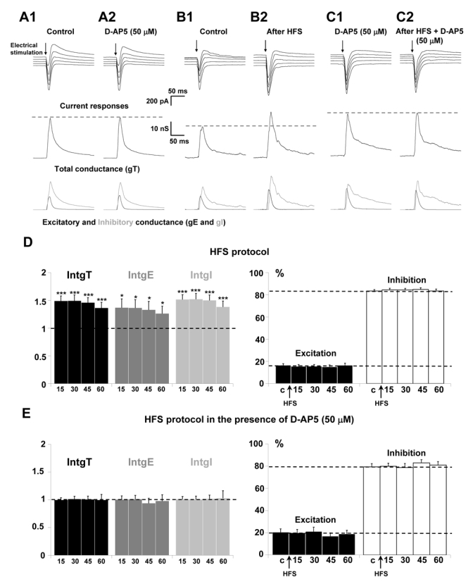
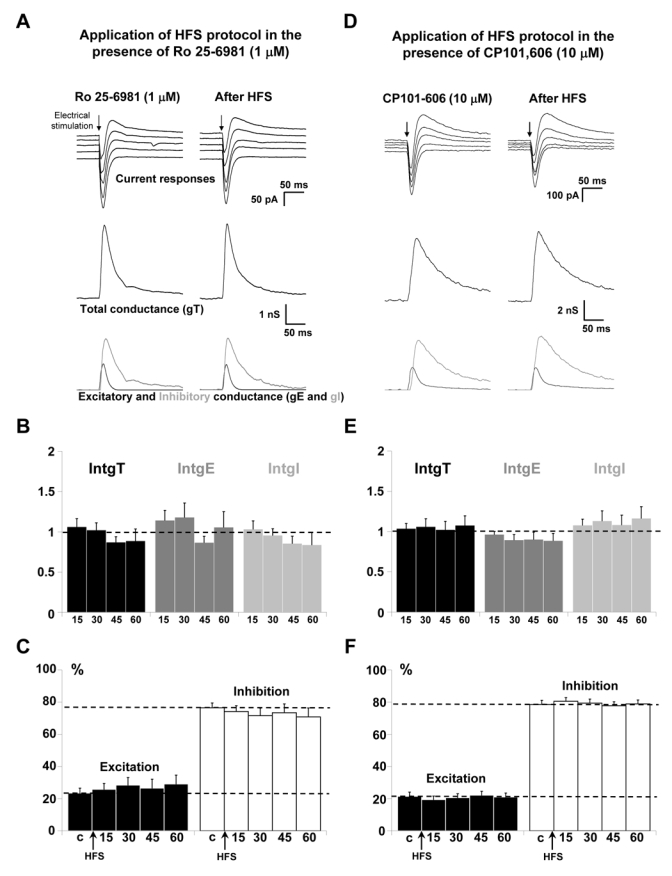
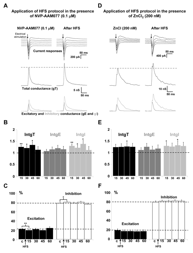
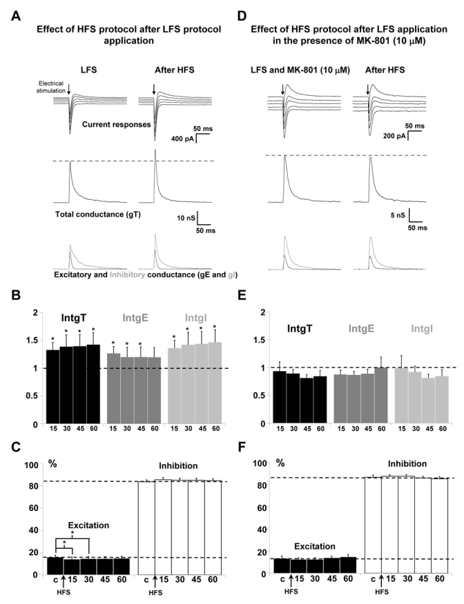
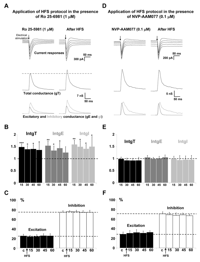
In the cortex, N-methyl-D-aspartate receptors (NMDARs) play a critical role in the control of synaptic plasticity processes. We have previously shown in rat visual cortex that the application of a high-frequency stimulation (HFS) protocol used to induce long-term potentiation in layer 2/3 leads to a parallel potentiation of excitatory and inhibitory inputs received by cortical layer 5 pyramidal neurones without changing the excitation/inhibition balance of the pyramidal neurone, indicating a homeostatic control of this parameter. We show here that the blockade of NMDARs of the neuronal network prevents the potentiation of excitatory and inhibitory inputs, and this result leaves open to question the role of the NMDAR isoform involved in the induction of long-term potentiation, which is actually being strongly debated. In postnatal day (P)18-23 rat cortical slices, the blockade of synaptic NR2B-containing NMDARs prevents the induction of the potentiation induced by the HFS protocol, whereas the blockade of NR2A-containing NMDARs reduced the potentiation itself. In P29-P32 cortical slices, the specific activation of NR2A-containing receptors fully ensures the potentiation of excitatory and inhibitory inputs. These results constitute the first report of a functional shift in subunit composition of NMDARs during the critical period (P12-P36), which explains the relative contribution of both NR2B- and NR2A-containing NMDARs in synaptic plasticity processes. These effects of the HFS protocol are mediated by the activation of synaptic NMDARs but our results also indicate that the homeostatic control of the excitation/inhibition balance is independent of NMDAR activation and is due to specialized recurrent interactions between excitatory and inhibitory networks.


Figures

Figure 1
Figure 1. Effects of NMDA receptor blockade on the potentiation of layer 5 pyramidal neurons inputs
(AC) Upper traces: current responses of a layer 5 pyramidal neuron to electrical stimulation (black arrow) applied in layer 2/3: (A1) under control conditions and (A2) 20 min after perfusion of 50 μM D-AP5; holding potentials scaled from − 75 (bottom trace) to − 55 mV (top trace, steps equal to 5 mV). (B1) Under control conditions and (B2) 15 min after HFS protocol; holding potentials scaled from − 70 (bottom trace) to − 50 mV (top trace, steps equal to 5 mV). (C1) 20 min after application of 50 μM D-AP5 and (C2) 15 min after HFS protocol in the presence of D-APS; holding potentials scaled from − 75 (bottom trace) to − 55 mV (top trace, steps equal to 5 mV). Medium traces: decomposition of the responses in total conductance change (gT). Lower traces: decomposition of gT in excitatory (gE, dark grey) and inhibitory (gI, light grey) conductance changes. Note that, for stimulation in layer 2/3 in the presence of D-AP5, no significant change of the response was observed. HFS protocol induced an increase of total, excitatory and inhibitory conductance changes which was prevented in the presence of D-AP5. (D) Left part: relative changes (compared to control) of intgT (black), intgE (dark grey) and intgI (light grey), after application of HFS protocol in layer 2/3 under control conditions (n = 26) (*** p < 0.001, ** p < 0.01 and * p < 0.05, t-test). Right part: relative contribution of excitatory and inhibitory conductances to the total conductance change, after HFS (c: control before HFS protocol). (E) Left part: relative changes (compared to control) of intgT (black), intgE (dark grey) and intgI (light grey), after HFS in layer 2/3 (n = 15) in the presence of 50 μM D-AP5. Right part: relative contribution of excitatory and inhibitory conductances to the total conductance change, after HFS in the presence of 50 μM D-AP5 (c: control before HFS protocol).
Figure 2
Figure 2. Effects of the blockade of NR2B-containing receptors on the potentiation of layer 5 pyramidal neurons inputs for P18–P23 old rats
(A) The left column shows representative recordings before HFS application and the right column 60 min after application of HFS in layer 2/3 in the presence of 1 μM Ro 25-6981. Upper traces: current responses of a layer 5 pyramidal neuron to electrical stimulation. Imposed membrane potential ranged for −55 to −75 mV. The amplitude of current responses was unchanged after HFS application in the presence of Ro 25-6981. Medium traces: decomposition of the responses in total conductance change (gT). Lower traces: decomposition of gT in excitatory and inhibitory conductance changes (gE, dark grey and gI, light grey). (B) Relative changes (compared to control) of intgT (black bars), intgE (dark grey bars) and intgI (light grey bars), after HFS in layer 2/3 (n = 12) in the presence of 1 μM Ro 25-6981. (C) Relative contribution of excitation and inhibition conductance changes to the total conductance change, after HFS in the presence of 1 μM Ro 25-6981 (c:control before HFS protocol). (D-E-F) Application of HFS protocol in the presence of 10 μM CP101,606 (n = 18). Legends are identical to those in A-B-C. No variations of total, excitatory and inhibitory conductance changes were observed after HFS (D-E) and the E/I ratio was unchanged (F).
Figure 3
Figure 3. Effects of the blockade of NR2A-containing receptors on the potentiation of layer 5 pyramidal neurons inputs for P18-P23 old rats
(A) The left column shows representative recordings before HFS application and the right column 60 min after application of HFS in layer 2/3 in the presence of 0.1 μM NVP-AAM077. Upper traces: current responses of a layer 5 pyramidal neuron to electrical stimulation. Imposed membrane potential ranged for −55 to −75 mV. The amplitudes of current responses were slightly increased after HFS application. Medium traces: decomposition of the responses in total conductance change (gT). Lower traces: decomposition of gT in excitatory and inhibitory conductance changes (gE, dark grey and gI, light grey). Total, excitatory and inhibitory conductance changes were all increased. (B) Relative changes (compared to control) of intgT (black bars), intgE (dark grey bars) and intgI (light grey bars), after HFS in layer 2/3 (n = 13) in the presence of 0.1 μM NVP-AAM077. (*** p < 0.001, ** p < 0.01 and * p < 0.05, t-test). (C) Relative contribution of excitation and inhibition conductance changes to the total conductance change, after HFS in the presence of 0.1 μM NVP-AAM077 (c:control before HFS protocol). (D-E-F) Application of HFS protocol in the presence of 200 nM ZnCl2 (n = 13). Legends are identical to those in A-B-C. (D) Imposed membrane potential ranged for −60 to −80 mV. (E-F) Total, excitatory and inhibitory conductance changes were increased after HFS.
Figure 4
Figure 4. Effect of synaptic NMDARs blockade on the potentiation of layer 5 pyramidal neurons inputs
(A) Left column: representative recordings after application of LFS protocol in layer 2/3. Right column: the LFS protocol was followed by an HFS protocol and recordings were done 60 min after the HFS protocol. Upper traces: current responses of a layer 5 pyramidal neuron to electrical stimulation. Imposed membrane potential ranged for −60 to −80 mV. The amplitudes of current responses were increased after HFS application. Medium traces: decomposition of the responses in total conductance change (gT). Lower traces: decomposition of gT in excitatory and inhibitory conductance changes (gE, dark grey and gI, light grey). Total, excitatory and inhibitory conductance changes were all increased. (B) Relative changes (compared to control) of intgT (black bars), intgE (dark grey bars) and intgI (light grey bars), after HFS in layer 2/3 (n = 14) after application of LFS protocol. (*** p < 0.001, ** p < 0.01 and * p < 0.05, t-test). (C) Relative contribution of excitation and inhibition conductance changes to the total conductance change, after application of LFS protocol, without MK 801. (c:control before HFS protocol). (D-E-F) Application of HFS after application of a LFS protocol in the presence of 10 μM MK-801 (n = 13). Legends are identical to those in A-B-C. No significant variations of total, excitatory and inhibitory conductance changes were observed after HFS.
Figure 5
Figure 5. Effects of the blockade of NR2B or NR2A-containing receptors on the potentiation of layer 5 pyramidal neurons inputs for P29-P32 old rats
(A) The left column shows representative recordings from the statistical analysis of n = 12 experiments before HFS application and the right column 60 min after application of HFS in layer 2/3 in the presence of 1 μM Ro 25-6981. Upper traces: current responses of a layer 5 pyramidal neuron to electrical stimulation. Imposed membrane potential ranged for −55 to −75 mV. The amplitude of current responses was increased after HFS application. Medium traces: decomposition of the responses in total conductance change (gT). Lower traces: decomposition of gT in excitatory and inhibitory conductance changes (gE, dark grey and gI, light grey). Total, excitatory and inhibitory conductance changes were all increased. (B) Relative changes (compared to control) of intgT (black bars), intgE (dark grey bars) and intgI (light grey bars), after HFS in layer 2/3 (n = 12) in the presence of 1 μM Ro 25-6981. (C) Relative contribution of excitation and inhibition conductance changes to the total conductance change, after HFS in the presence of 1 μM Ro 25-6981 (c:control before HFS protocol). (D-E-F) Application of HFS protocol in the presence of 0.1 μM NVP-AAM077 (n = 13). Legends are identical to those in A-B-C. No variations of total, excitatory and inhibitory conductance changes were observed after HFS (D-E) and the E/I ratio was unchanged (F).

Similar articles

Cited by

References

    1. Abraham WC, Huggett A. Induction and reversal of long-term potentiation by repeated high-frequency stimulation in rat hippocampal slices. Hippocampus. 1997;7:137–145. - PubMed
    1. Amar M, Perin-Dureau F, Neyton J. High-affinity Zn block in recombinant N-methyl-D-aspartate receptors with cysteine substitutions at the Q/R/N site. Biophys J. 2001;81:107–116. - PMC - PubMed
    1. Anderson JS, Carandini M, Ferster D. Orientation tuning of input conductance, excitation, and inhibition in cat primary visual cortex. J Neurophysiol. 2000;84:909–926. - PubMed
    1. Auberson YP, Allgeier H, Bischoff S, Lingenhoehl K, Moretti R, Schmutz M. 5-Phosphonomethylquinoxalinediones as competitive NMDA receptor antagonists with a preference for the human 1A/2A, rather than 1A/2B receptor composition. Bioorg Med Chem Lett. 2002;12:1099–1102. - PubMed
    1. Babb TL, Mikuni N, Najm I, Wylie C, Olive M, Dollar C, MacLennan H. Pre- and postnatal expressions of NMDA receptors 1 and 2B subunit proteins in the normal rat cortex. Epilepsy Res. 2005;64:23–30. - PubMed

MeSH terms