. 2007 Jul;26(2):439-45.
doi: 10.1111/j.1460-9568.2007.05686.x.

The chemical component of the mixed GF-TTMn synapse in Drosophila melanogaster uses acetylcholine as its neurotransmitter

Affiliations

The chemical component of the mixed GF-TTMn synapse in Drosophila melanogaster uses acetylcholine as its neurotransmitter

Marcus J Allen et al. Eur J Neurosci. 2007 Jul.

Abstract

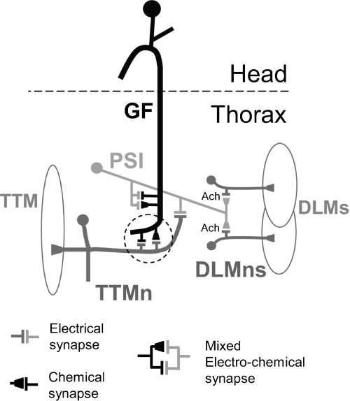
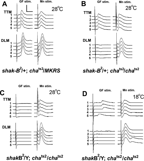
The largest central synapse in adult Drosophila is a mixed electro-chemical synapse whose gap junctions require the product of the shaking-B (shak-B) gene. Shak-B(2) mutant flies lack gap junctions at this synapse, which is between the giant fibre (GF) and the tergotrochanteral motor neuron (TTMn), but it still exhibits a long latency response upon GF stimulation. We have targeted the expression of the light chain of tetanus toxin to the GF, to block chemical transmission, in shak-B(2) flies. The long latency response in the tergotrochanteral muscle (TTM) was abolished indicating that the chemical component of the synapse mediates this response. Attenuation of GAL4-mediated labelling by a cha-GAL80 transgene, reveals the GF to be cholinergic. We have used a temperature-sensitive allele of the choline acetyltransferase gene (cha(ts2)) to block cholinergic synapses in adult flies and this also abolished the long latency response in shak-B(2) flies. Taken together the data provide evidence that both components of this mixed synapse are functional and that the chemical neurotransmitter between the GF and the TTMn is acetylcholine. Our findings show that the two components of this synapse can be separated to allow further studies into the mechanisms by which mixed synapses are built and function.


Figures

F<sc>ig</sc>. 1
Fig. 1
Schematic representing the known synaptic connections of the GFS. For simplicity only one side of the bilateral circuit is shown. The GF makes mixed electrochemical synapses with the PSI and with the TTMn in the thoracic ganglia. The GF-TTMn synapse is circled with a dotted line. The PSI synapses with the DLMns via cholinergic chemical synapses. The PSI synapses with five DLMns, but only two are indicated for clarity. PSI and TTMn are also electrically coupled. Adapted from Allen et al. (2006).
F<sc>ig</sc>. 2
Fig. 2
Expression of tetanus toxin in the GF abolishes the TTM response in shak-B2 mutants. Responses in the TTM and a DLM are shown when individual flies were given a single brain stimulus or ten brain stimuli at either 100 or 250 Hz. (A–C) responses in a shak-B2/+ c17/UAS-IMPTNT control fly show wild-type latencies and following frequencies at 100 and 250 Hz including the DLM not following 1:1 at 250 Hz (*) due to the failure of the PSI-DLMns synapses (Tanouye & Wyman, 1980). (D–F) responses in a shak-B2/Y c17/UAS-IMPTNT fly showing no output to DLM and a long latency response and poor following in TTM at both frequencies. (G–I) responses in a shak-B2/Y c17/UAS-TNT fly show no responses in either TTM or DLM upon stimulation but increased spontaneous activity (marked with arrows). Vertical scale bars, 50 mV for all traces; horizontal, 1 ms for response latencies, 10 ms for following at 100 Hz, and 4 ms for following at 250 Hz.
F<sc>ig</sc>. 3
Fig. 3
NMJ function is unaffected by the shak-B2 mutation or expression of tetanus toxin using the c17 line. (A) Schematic showing the positions of the stimulating and recording electrodes for either GF or motorneuron stimulation. (B) Responses in TTM and DLM to a single stimulus, or ten stimuli at 250 Hz, in the brain (GF stimulation) or the thorax (Mn stimulation) from a shak-B2/Y UAS-TNT/CyO fly. (C) Responses in TTM and DLM to a single stimulus, or ten stimuli at 250 Hz, in the brain (GF stimulation) or the thorax (Mn stimulation) from a shak-B2/Y c17/UAS-TNT fly. In (B) and (C) Mn stimulation always resulted in a muscle response to every stimulus.
F<sc>ig</sc>. 4
Fig. 4
The GF is a cholinergic neuron. Dissected adult nervous systems stained for LacZ. (A) UAS-lacZ; A307 control preparation showing distinct staining in the GFs (*) and a few other cells in the brain and ventral nerve cord including a cell that lies just ventral to each GF (arrowhead). Inset is a higher power view of a cervical connective through which the labelled GFs can be easily identified. (B) UAS-lacZ; A307; cha3.3kb-GAL80 preparation. Note the lack of staining in the GFs but the presence of staining in the small ventral cell (arrowhead) that is in the position to be a cell body of a giant commissural interneuron. Inset higher power view of a cervical connective shows the GFs to be present but unlabelled. Scale bar, 50 µm; 25 µm for insets.
F<sc>ig</sc>. 5
Fig. 5
The response in the TTM is blocked in shak-B2; chats2 double-mutants at the restrictive temperature. Traces from the TTM and DLM of individual flies given five stimuli (1–5) at 1 Hz. (A) shak-B2/+ chats2/MKRS control female showing WT responses upon GF stimulation (GF stim) at 28 °C. (B) shak-B2/+ chats2/chats2 female showing a normal response in TTM and a loss of the DLM response at 28 °C. (C) shak-B2/Y; chats2/chats2 male showing no responses in DLM and a loss of responses in TTM at 28 °C. (D) shak-B2/Y; chats2/chats2 male showing no responses in DLM but responses in TTM at 18 °C. In all cases, individuals showed responses in both muscles upon thoracic stimulation (Mn stim.).Vertical scale bar, 50 mV; horizontal scale bar, 2 ms.

Similar articles

Cited by

References

    1. Allen MJ, Drummond JA, Moffat KG. Development of the giant fiber neuron of Drosophila melanogaster. JCompNeurol. 1998;397:519–531. - PubMed
    1. Allen MJ, Godenschwege TA, Tanouye MA, Phelan P. Making an escape: development and function of the Drosophila giant fibre system. SeminCellDevBiol. 2006;17:31–41. - PubMed
    1. Allen MJ, Shan X, Caruccio P, Froggett SJ, Moffat KG, Murphey RK. Targeted expression of truncated glued disrupts giant fiber synapse formation in Drosophila. JNeurosci. 1999;19:9374–9384. - PMC - PubMed
    1. Allen MJ, Shan XL, Murphey RK. A role for Drosophila Drac1 in neurite outgrowth and synaptogenesis in the giant fiber system. MolCellNeurosci. 2000;16:754–765. - PubMed
    1. Baird DH, Schalet AP, Wyman RJ. The Passover locus in Drosophila melanogaster: complex complementation and different effects on the giant fiber neural pathway. Genetics. 1990;126:1045–1059. - PMC - PubMed

Publication types

MeSH terms

LinkOut - more resources