. 2007 Jul;26(2):510-22.
doi: 10.1111/j.1460-9568.2007.05678.x.

Deafness alters auditory nerve fibre responses to cochlear implant stimulation

Affiliations

Deafness alters auditory nerve fibre responses to cochlear implant stimulation

David J Sly et al. Eur J Neurosci. 2007 Jul.

Abstract

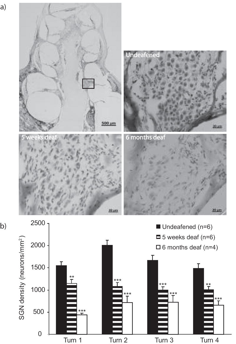
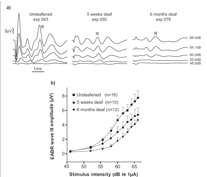
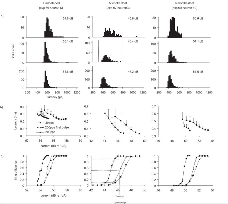
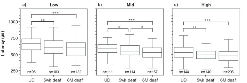
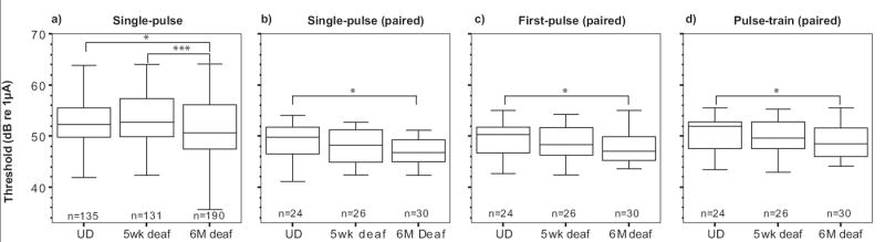
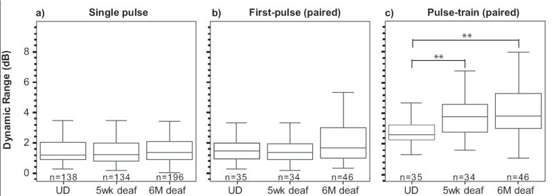
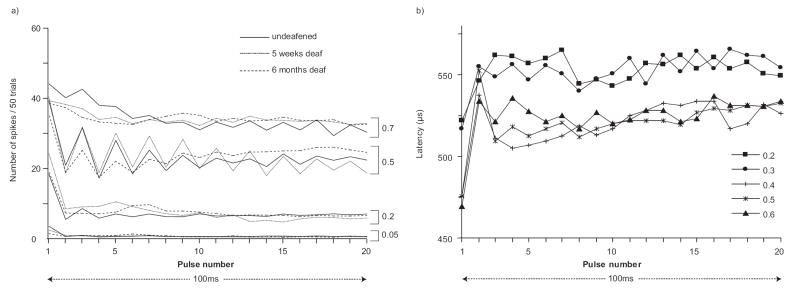
Here we characterized the relationship between duration of sensorineural hearing loss and the response of the auditory nerve to electrical stimulus rate. Electrophysiological recordings were made from undeafened guinea pigs and those ototoxically deafened for either 5 weeks or 6 months. Auditory neuron survival decreased significantly with the duration of deafness. Extracellular recordings were made from auditory nerve fibres responding to biphasic, charge-balanced current pulses delivered at rates of 20 and 200 pulses/s via a monopolar scala tympani stimulating electrode. The response to 20 pulses/s electrical stimulation of the deafened cochlea exhibited a decrease in spike latency, unaltered temporal jitter and unaltered dynamic range (of nerve firing rate against stimulus current), and a reduction in threshold after 6 months of deafness. The response to a 200-pulse/s stimulus was similar except that the dynamic range was greater than with 20 pulses/s and was also greater in deafened animals than in undeafened animals. Deafness and pulse rate are related; in deaf animals spike recovery appears to be complete between successive stimulus pulses at a low rate (20 pulses/s), but incomplete between pulses at a moderate pulse rate (200 pulses/s). These results suggest that changes in the function of individual auditory nerve fibres after deafness may affect clinical responses during high-rate stimulation such as that used in contemporary speech processing strategies, but not during lower rate stimulation such as that used to record evoked potentials.


Figures

Fig 1
Fig 1
a) Photomicrographs showing the loss of spiral ganglion neurons within Rosenthal’s canal that occurs after chronic sensorineural hearing loss. The rectangle on the upper left image indicates the region of turn 2 of the cochlea from which all other images are taken. b) Spiral ganglion neuron densities calculated at each cochlear turn. Turn 1 is the most basal and turn 4 is the most apical. Each bar represents the mean±SEM density averaged across the number of animals indicated in the figure and across five sections per animal. Asterisks represent ANOVA comparisons between undeafened and deafened groups, **p<.01, ***p<0.001.
Fig 2
Fig 2
a) Example traces of electrically-evoked auditory brainstem responses (EABRs) from one animal in each of the treatment groups. The stimulus current was delivered via a bipolar cochlear electrode configuration at 33 pps using a 50μs biphasic pulse. The amplitude of wave III increased with stimulus amplitude and was lower in the deafened animals. b) EABR growth functions of peak wave III amplitude plotted against stimulus current. Each data point shows the mean±SEM of the peak amplitude of wave III at each current level, across the number of animals indicated in the figure. EABR growth functions were reduced in amplitude after five weeks and six months of deafness. Statistical comparisons at the maximum stimulus current (66dB re 1 μA) revealed the median wave III amplitudes from the six month deafened group were significantly lower than those of the undeafened group, U, z=-2.284, p=0.022). Although not significant, the mean wave III amplitudes from the five week deafened group were also lower than those of the undeafened group.
Fig 3
Fig 3
Example ANF data from undeafened, 5-week-deafened and 6-month-deafened guinea pigs is shown in the three panels. a) Post-stimulus time histograms at 0.5 firing efficiency (200-pps). Spontaneous activity is present as scattered events through the time-course of the recording. The dashed vertical lines mark the time interval over which these responses were windowed (see main text). b) Average latency±standard deviation (jitter) across trials plotted against stimulus current. c) Input-output (IO) functions of firing efficiency against stimulus current calculated from the same neuron for stimuli at both 20 and 200 pulses per second.
Fig 4
Fig 4
Latency decreased with sensorineural hearing loss and firing efficiency. Auditory nerve fibre (ANF) latencies are shown calculated at a) low (0-0.2) b) mid (0.4-0.6) and c) high firing (0.8-1.0) efficiencies during single-pulse stimulation. Each box plot represents pooled data from the number of ANFs indicated. The upper and lower limits of each box indicate the 25th-75th percentiles respectively, with the median indicated inside each box. The lower and upper range bars outside each box indicate the 5th-95th percentiles respectively. The effect of hearing status is compared within each panel. UD=undeafened. U, *p<0.05, **p<0.01, ***p<0.001.
Fig 5
Fig 5
Threshold decreased with increasing duration of sensorineural hearing loss. The threshold was defined as the stimulus intensity required to evoke 0.5 firing efficiency response. Responses were calculated from an integrated Gaussian curve fitted to input-output functions resulting from a) 20pps and paired data showing only neurons where responses to both 20pps and 200pps stimuli were recorded for b) 20pps response c) the response to the first pulse of the pulse-train stimuli and d) 200pps response. The effect of hearing status is compared within each panel. UD=undeafened. U, *p<0.05, ***p<0.001.
Fig 6
Fig 6
Dynamic range of auditory nerve fibres to electrical stimulation is unaltered by sensorineural hearing loss during a) stimulation at 20 pulses per second and in response to b) the first pulse of 200pps pulse-train stimulation but c) increases with sensorineural hearing loss during 200 pulses per second stimulation. UD=undeafened. U, **p<0.01.
Fig 7
Fig 7
a) Post-stimulus time histograms in response to the 200pps pulse-train stimulation and b) the mean latencies in response to each stimulus pulse of the 200pps pulse-train. These plots were obtained by pooling data across all neurons in each group (UD= undeafened, five week and six month deaf) within specific ranges of firing efficiency (see methods). The plot reveals accommodation of the response, and a saw-tooth-like pattern of response. b) The latency plots also possess a saw-tooth pattern, with the shorter latencies corresponding to the pulses with the higher firing efficiencies.
Fig 8
Fig 8
Two examples of bursting exhibited during single-pulse stimulation. Each bin represents one stimulus pulse (or “trial”). Pulses eliciting a spike were allocated a count of one, otherwise zero was recorded. The inter-pulse interval was 50 ms.

References

    1. Black RC, Clark GM, O’Leary SJ, Walters C. Intracochlear electrical stimulation of normal and deaf cats investigated using brainstem response audiometry. Acta Otolaryngol Suppl. 1983;399:5–17. - PubMed
    1. Blamey PJ, Ardnt P, Bergeron G, Brimacombe J, Facer G, Larky J, Lindstrom B, Nedzelski J, Peterson J, Peterson A, Shipp D, Staller S, Whitford L. Factors Affecting Auditory Performance of Postlinguistically Deaf Adults Using Cochlear Implants. Audiol Neurootol. 1996;1:293–306. - PubMed
    1. Bruce IC, Irlicht LS, White MW, O’Leary SJ, Javel E, Clark GM. A stochastic model of the electrically stimulated auditory nerve: single-pulse response. IEEE Trans Biomed Eng. 1999;46:617–629. - PubMed
    1. Dodson HC, Mohuiddin A. Response of spiral ganglion neurones to cochlear hair cell destruction in the guinea pig. J Neurocytol. 2000;29:525–537. - PubMed
    1. Friedman L. Ph.D. Dissertation. University Of Melbourne; Australia: 2002. A retrospective study on the outcomes of cochlear implants in patients with otosclerosis.

Publication types