

Computerized tomography myelography with coronal and oblique coronal view for diagnosis of nerve root avulsion in brachial plexus injury
- PMID: 17651476
- PMCID: PMC1947985
- DOI: 10.1186/1749-7221-2-16
Computerized tomography myelography with coronal and oblique coronal view for diagnosis of nerve root avulsion in brachial plexus injury
Abstract
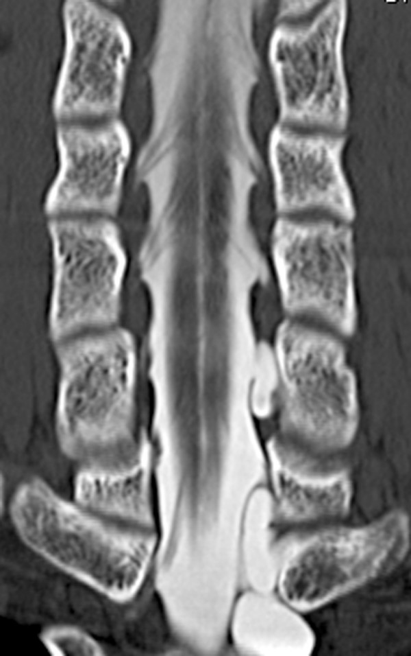
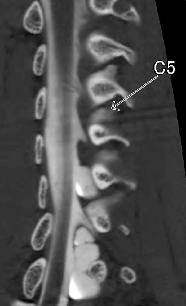
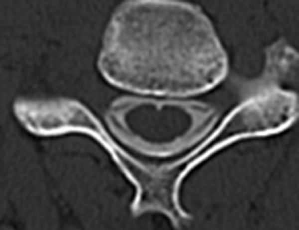
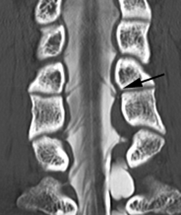
Background: The authors describe a new computerized tomography (CT) myelography technique with coronal and oblique coronal view to demonstrate the status of the cervical nerve rootlets involved in brachial plexus injury. They discuss the value of this technique for diagnosis of nerve root avulsion compared with CT myelography with axial view.
Methods: CT myelography was performed with penetration of the cervical subarachnoid space by the contrast medium. Then the coronal and oblique coronal reconstructions were created. The results of CT myelography were evaluated and classified with presence of pseudomeningocele, intradural ventral nerve rootlets, and intradural dorsal nerve rootlets. The diagnosis was by extraspinal surgical exploration with or without spinal evoked potential measurements and choline acetyl transferase activity measurement in 25 patients and recovery by a natural course in 3 patients. Its diagnostic accuracy was compared with that of CT myelography with axial view, correlated with surgical findings or a natural course in 57 cervical roots in 28 patients.
Results: Coronal and oblique coronal views were superior to axial views in visualization of the rootlets and orientation of the exact level of the root. Sensitivity and specificity for coronal and oblique coronal views of unrecognition of intradural ventral and dorsal nerve root shadow without pseudomeningocele in determining pre-ganglionic injury were 100% and 96%, respectively. There was no statistically significant difference between coronal and oblique coronal views and axial views.
Conclusion: The information by the coronal and oblique coronal slice CT myelography enabled the authors to assess the rootlets of the brachial plexus and provided valuable data for helping to decide whether to proceed with exploration, nerve repair, primary reconstruction.
Figures
References
-
- Doi K, Otsuka K, Okamoto Y, Fujii H, Hattori Y, Baliarsing AS. Cervical nerve root avulsion in brachial plexus injuries: magnetic resonance imaging classification and comparison with myelography and computerized tomography myelography. J Neurosurg. 2002;2:277–284. - PubMed
-
- Carvalho GA, Nikkhah G, Matthies C, Penkert G, Samii M. Diagnosis of root avulsions in traumatic brachial plexus injuries: value of computerized tomography myelography and magnetic resonance imaging. J Neurosurg. 1997;2:69–76. - PubMed
-
- Hattori Y, Doi K, Dhawan V, Ikeda K, Kaneko K, Ohi R. Choline acetyltransferase activity and evoked spinal cord potentials for diagnosis of brachial plexus injury. J Bone Joint Surg Br. 2004;2:70–73. - PubMed
LinkOut - more resources
Full Text Sources
Other Literature Sources