. 2007 Jul 25:7:137.
doi: 10.1186/1471-2407-7-137.

PI3K activation is associated with intracellular sodium/iodide symporter protein expression in breast cancer

Affiliations

PI3K activation is associated with intracellular sodium/iodide symporter protein expression in breast cancer

Katherine A B Knostman et al. BMC Cancer. .

Abstract

Background: The sodium/iodide symporter (NIS) is a membrane glycoprotein mediating active iodide uptake in the thyroid gland and is the molecular basis for radioiodide imaging and therapeutic ablation of thyroid carcinomas. NIS is expressed in the lactating mammary gland and in many human breast tumors, raising interest in similar use for diagnosis and treatment. However, few human breast tumors have clinically evident iodide uptake ability. We previously identified PI3K signaling as important in NIS upregulation in transgenic mouse models of breast cancer, and the PI3K pathway is commonly activated in human breast cancer.

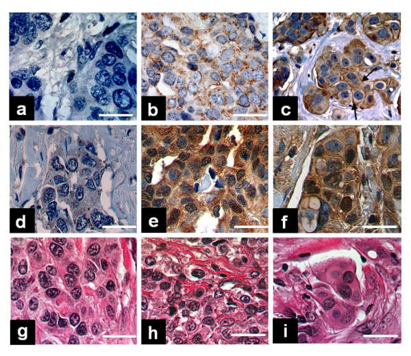
Methods: NIS expression, subcellular localization, and function were analyzed in MCF-7 human breast cancer cells and MCF-7 cells stably or transiently expressing PI3K p110alpha subunit using Western blot of whole cell lysate, cell surface biotinylation Western blot and immunofluorescence, and radioiodide uptake assay, respectively. NIS localization was determined in a human breast cancer tissue microarray using immunohistochemical staining (IHC) and was correlated with pre-existing pAkt IHC data. Statistical analysis consisted of Student's t-test (in vitro studies) or Fisher's Exact Test (in vivo correlational studies).

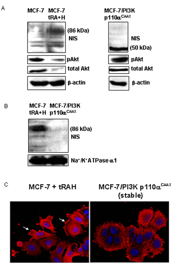
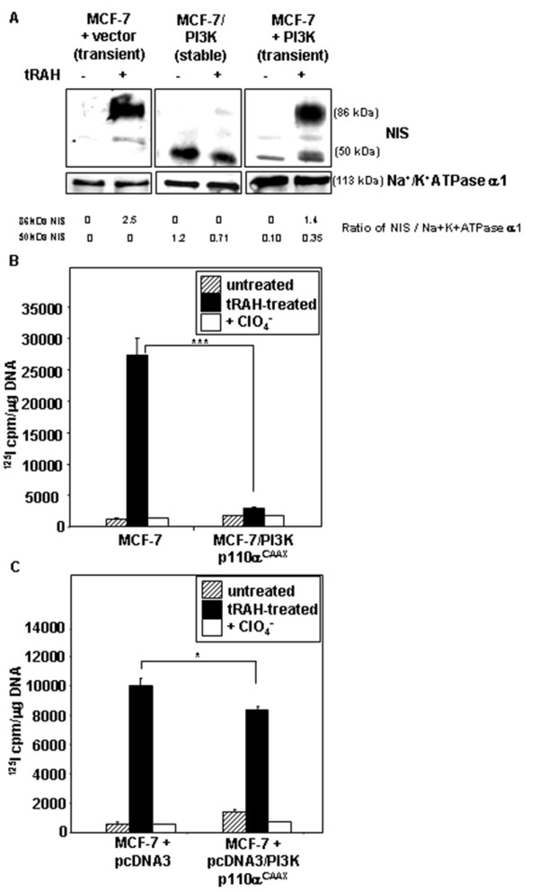
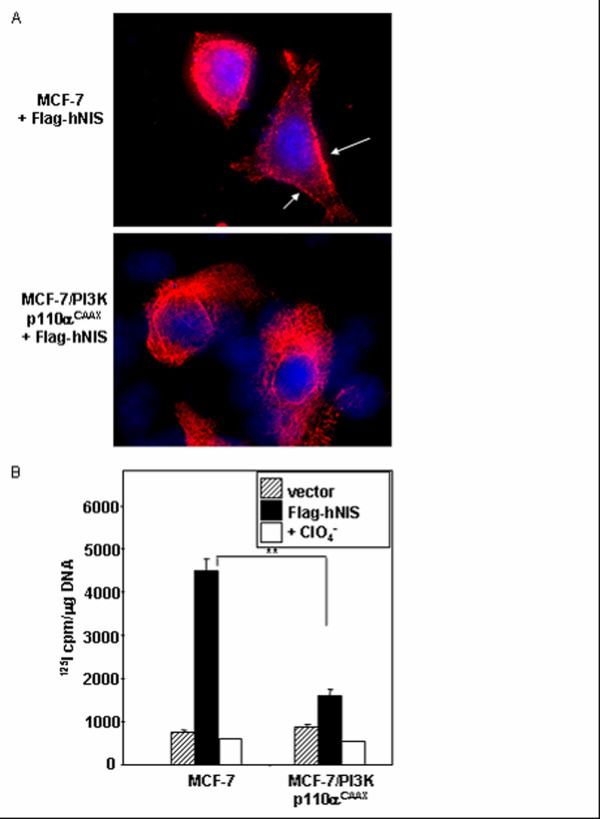
Results: In this study, we demonstrate that PI3K activation in MCF-7 human mammary carcinoma cells leads to expression of underglycosylated NIS lacking cell surface trafficking necessary for iodide uptake ability. PI3K activation also appears to interfere with cell surface trafficking of exogenous NIS as well as all-trans retinoic acid-induced endogenous NIS. A correlation between NIS expression and upregulation of PI3K signaling was found in a human breast cancer tissue microarray.

Conclusion: Thus, the PI3K pathway likely plays a major role in the discordance between NIS expression and iodide uptake in breast cancer patients. Further study is warranted to realize the application of NIS-mediated radioiodide ablation in breast cancer.


Figures

Figure 1
Figure 1
PI3K activation induces underglycosylated intracellular NIS protein expression in MCF-7 cells. (A) immunoblotting of membrane-enriched lysates from MCF-7 cells with and without tRAH treatment and MCF-7/PI3K p110αCAAX stable clones using human NIS (hNIS) antibody. The specificity of NIS antibody was confirmed by conversion of the 86 kDa NIS band to a single 50 kDa band upon PNGase F deglycosylation (data not shown). Faint non-specific bands of 60 and 70 kDa are present. PI3K activation was assessed by total and phospho-Akt immunoblotting of total cell lysates. Actin was used as a loading control. (B) Immunoblotting of the surface protein fraction from MCF-7 cells treated with tRAH and MCF-7/PI3K p110αCAAX stable clones using hNIS antibody. The surface fraction was isolated by cell surface biotinylation and avidin pull-down. Na+/K+ATPase was used as a loading control. (C) tRAH-treated MCF-7 cells and MCF-7/PI3K p110αCAAX cells were labeled with hNIS antibody followed by CyTM3-conjugated secondary antibody (red color) and DAPI nuclear stain (blue) for immunofluorescent microscopy. Parental MCF-7 cells and secondary antibody only controls were utilized, but are not shown. Magnification = 63×.
Figure 2
Figure 2
Stable or acute PI3K activation decreases tRAH-induced glycosylated NIS protein expression and NIS-mediated radioactive iodide uptake in MCF-7 cells. (A) 48 hours of tRAH treatment induces fully glycosylated NIS protein in MCF-7 cells transiently transfected with empty vector. Underglycosylated 50 kDa NIS is dominant in MCF-7/PI3K p110αCAAX cells, which minimally respond to tRAH treatment. Transient expression of PI3K p110αCAAX also induces underglycosylated NIS protein expression with a modest decrease in the tRAH-induced fully glycosylated NIS form. (B) tRAH treatment cannot induce NIS function in MCF-7/PI3K p110αCAAX cells. A small increase in basal NIS-mediated radioactive iodide uptake is noted in MCF-7/PI3K p110αCAAX stable clones versus parental MCF-7 cells. Cells were treated with tRAH for 48 hours followed by 125I uptake assay. Perchlorate (ClO4-) is a specific inhibitor of NIS function. ***p < .0001. (C) Acute expression of activated PI3K p110α decreases tRAH-induced NIS function in MCF-7 cells. A modest increase in basal NIS-mediated radioactive iodide uptake is present in MCF-7 cells transiently transfected with PI3K p110αCAAX versus vector-only controls. Cells were transiently transfected with pcDNA3/PI3K p110αCAAX or empty vector for 6 hours, and then treated with tRAH for 42 hours prior to 125I uptake assay. *p < .05.
Figure 3
Figure 3
PI3K activation interferes with cell surface trafficking and radioiodide uptake conferred by exogenous Flag-tagged hNIS gene expression. (A) MCF-7 and MCF-7/PI3K p110αCAAX cells were transiently transfected with pcDNA3/Flag-hNIS and subjected to Flag immunofluorescence 48 hours later. While Flag-hNIS is detectable on the cell surface in parental MCF-7 cells (arrows), it is retained intracellularly in MCF-7/PI3K p110αCAAX cells. Magnification = 63×. (B) MCF-7 and MCF-7/PI3K p110αCAAX cells were transiently transfected with pcDNA3/Flag-hNIS and subjected to 125I uptake assay 48 hours later. A nearly 4-fold increase in 125I uptake resulting from exogenous NIS expression is present in parental MCF-7 cells as compared to MCF-7/PI3K p110αCAAX. **p < .001.
Figure 4
Figure 4
NIS is expressed in 80% of human breast tumors in a tissue microarray with primarily intracellular localization and positive correlation with pAkt expression. (A-C) NIS immunohistochemical staining in three representative breast cancer specimens. (A) NIS-negative breast tumor. (B) Breast tumor with intracellular NIS localization. (C) Breast tumor with both intracellular and plasma membrane-localized NIS protein (arrows). (D-F) Immunohistochemical staining for pAkt in the three breast tumors shown in A-C. (G-I) H & E staining demonstrating the histopathology of the three breast tumors shown in A-C. Bar = 20 μM.

Similar articles

Cited by

References

    1. American Cancer Society, Breast Cancer http://www.cancer.org
    1. Taurog A. Hormone Synthesis. In: Braverman LL, Utiger RD, editor. Werner and Ingbar's The Thyroid: A Fundamental and Clinical Text. 7. Philadelphia, PA: J.B. Lippincott Co; 1996. pp. 47–81.
    1. Cotran RS, Kumar V, Collins T. The Endocrine System. In: Cotran RS, Kumar V, Collins T, editor. Robbins Pathologic Basis of Disease. 6. Philadelphia, PA: W.B. Saunders Company; 1999. pp. 1142–1147.
    1. Sherman SI. Thyroid Carcinoma. Lancet. 2003;361:501–511. doi: 10.1016/S0140-6736(03)12488-9. - DOI - PubMed
    1. Tazebay UH, Wapnir IL, Levy O, Dohan O, Zuckier LS, Zhao QH, Deng HF, Amenta PS, Fineberg S, Pestell RG, Carrasco N. The mammary gland iodide symporter is expressed during lactation and in breast cancer. Nat Med. 2000;6:871–878. doi: 10.1038/78630. - DOI - PubMed

Publication types