. 2007;8(7):R148.
doi: 10.1186/gb-2007-8-7-r148.

Co-localization of CENP-C and CENP-H to discontinuous domains of CENP-A chromatin at human neocentromeres

Affiliations

Co-localization of CENP-C and CENP-H to discontinuous domains of CENP-A chromatin at human neocentromeres

Alicia Alonso et al. Genome Biol. 2007.

Abstract

Background: Mammalian centromere formation is dependent on chromatin that contains centromere protein (CENP)-A, which is the centromere-specific histone H3 variant. Human neocentromeres have acquired CENP-A chromatin epigenetically in ectopic chromosomal locations on low-copy complex DNA. Neocentromeres permit detailed investigation of centromeric chromatin organization that is not possible in the highly repetitive alpha satellite DNA present at endogenous centromeres.

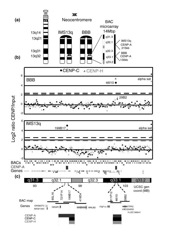
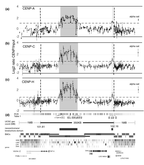
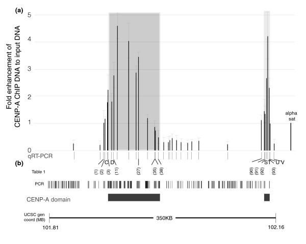
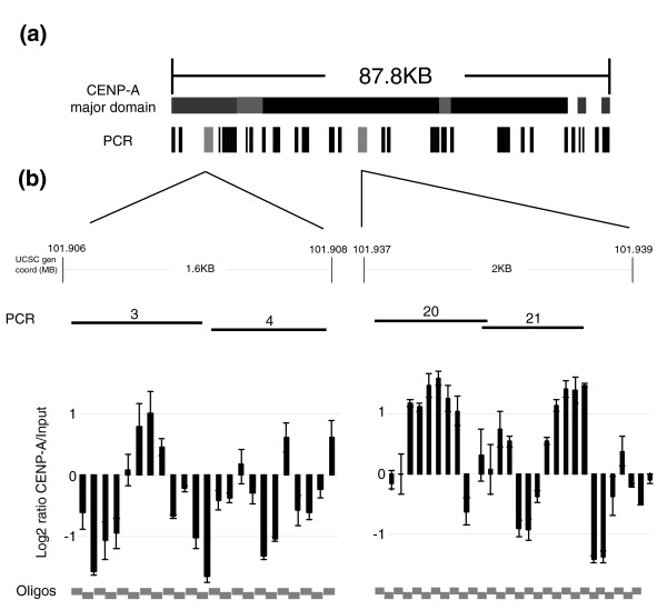
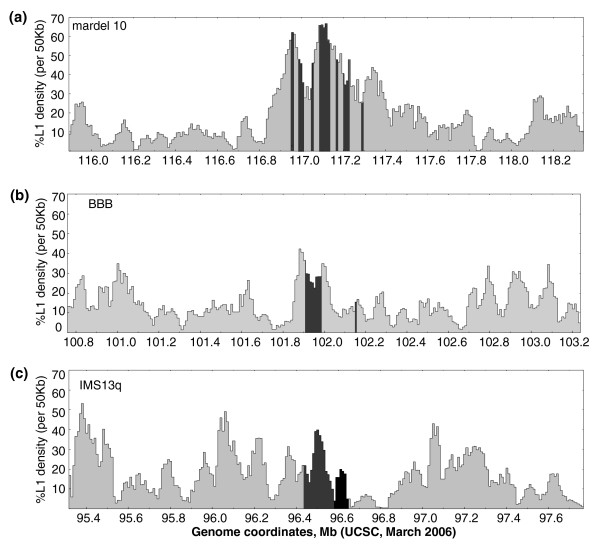
Results: We have examined the distribution of CENP-A, as well as two additional centromeric chromatin-associated proteins (CENP-C and CENP-H), across neocentromeric DNA using chromatin immunoprecipitation (ChIP) on CHIP assays on custom genomic microarrays at three different resolutions. Analysis of two neocentromeres using a contiguous bacterial artificial chromosome (BAC) microarray spanning bands 13q31.3 to 13q33.1 shows that both CENP-C and CENP-H co-localize to the CENP-A chromatin domain. Using a higher resolution polymerase chain reaction (PCR)-amplicon microarray spanning the neocentromere, we find that the CENP-A chromatin is discontinuous, consisting of a major domain of about 87.8 kilobases (kb) and a minor domain of about 13.2 kb, separated by an approximately 158 kb region devoid of CENPs. Both CENP-A domains exhibit co-localization of CENP-C and CENP-H, defining a distinct inner kinetochore chromatin structure that is consistent with higher order chromatin looping models at centromeres. The PCR microarray data suggested varying density of CENP-A nucleosomes across the major domain, which was confirmed using a higher resolution oligo-based microarray.

Conclusion: Centromeric chromatin consists of several CENP-A subdomains with highly discontinuous CENP-A chromatin at both the level of individual nucleosomes and at higher order chromatin levels, raising questions regarding the overall structure of centromeric chromatin.


Figures

Figure 1
Figure 1
Genomic microarray analysis of CENP-C and CENP-H binding domains in two independent 13q32/33 neocentromeres. (a) Ideogrammatic representation of the two neocentric chromosomes analyzed. From left to right: a normal chromosome 13, the invdup13q21 in IMS13q with a neocentromere in band 13q32, and the invdup13q21 in BBB with a neocentromere in band 13q33.1. An expansion of the 13q31.3 to 13q33.2 area included in the bacterial artificial chromosome (BAC) CHIP is shown. The position and size of each previously mapped centromere protein (CENP)-A domain from Alonso and coworkers [32] are indicated. (b) DNA obtained from chromatin immunoprecipitation (ChIP) using antibodies to CENP-C (circles) and CENP-H (triangles) from cell lines BBB and IMS13q was hybridized to a contiguous BAC microarray spanning 14 megabases (Mb) from 13q31.3 to 13q33.2. Shown across the bottom of the graph is the tiling path of the unique sequenced regions for each BAC, the previously determined CENP-A domains [32] in cell lines BBB and IMS13q, and the genes in the region. Three independent biologic replicates were performed for each ChIP from each cell line, and the scale normalized mean log2 Cy-5:Cy-3 intensity ratios (ChIP to input) with standard error (SE) were plotted on the y-axis for each BAC. Positive intensity ratios were identified as those that were at least three times the standard deviation (SD) from the experimental mean (gray or black dashed lines; see Materials and methods). For cell line BBB, CENP-C ChIP, the experimental mean was 0 ± 0.82 SD. Positive values ≥ 2.5 (black dashed line) were as follows: alpha sat = 6.42 ± 0.39 SE and BAC RP11-46I10 = 4.66 ± 0.92 SE. BAC RP11-29B2 was slightly increased (1.18 ± 1.2 SE) but not statistically significantly. All other BACs ranged from -1.1 to ≤ 0.96. For cell line BBB, CENP-H ChIP, the experimental mean was -0.02 ± 0.75 SD. Positive values ≥ 2.2 (grey dashed line) were as follows: alpha sat = 4.92 ± 1.86 SE and BAC RP11-46I10 = 5.57 ± 0.77 SE. BAC RP11-29B2 was slightly increased (1.58 ± 0.71 SE) but not statistically significantly. All other BACs ranged from -1.27 to ≤ 1.03. For cell line IMS13q, CENP-C ChIP, the experimental mean was 0 ± 0.84 SD. Positive values ≥ 2.5 (black dashed line) were as follows: alpha sat = 5.26 ± 0.38 SE and BAC RP11-199B17 = 4.95 ± 0.86 SE. All other BACs ranged from -1.7 to ≤ 0.93. For cell line IMS13q, CENP-H, the experimental mean was 0.00 ± 0.64 SD. Positive values ≥ 1.9 (grey dashed line) were as follows: alpha sat = 2.63 ± 1.03 SE and BAC RP11-199B17 = 3.95 ± 1.06 SE. All other BACs ranged from -1.17 to ≤ 1.13. (c) Expansion of BAC map in regions that are positive for CENP-C and CENP-H in each neocentromere examined, showing BAC names and overlaps, the genes, and the previously determined CENP-A domains. For cell line BBB, the CENP-A, CENP-C, and CENP-H were mapped to the identical BACs (negative for RP11-811P12, strongly positive for BAC 46I10, and weakly positive for 29B2). For cell line IMS13q, the CENP-A mapped to two contiguous BACs (RP11-721F4 and RP11-199B17), whereas CENP-C and CENP-H mapped only to one BAC (RP11-199B17).
Figure 2
Figure 2
The BBB neocentromere contains a major and a minor centromere chromatin domain. DNA obtained from chromatin immunoprecipitation (ChIP) using antibodies to CENP-A, CENP-C, and CENP-H from cell line BBB was hybridized to a custom made microarray containing 257 unique polymerase chain reaction (PCR) fragments. Three independent biological replicates were performed for each antibody, and the scale normalized mean log2 Cy-5:Cy-3 intensity ratios (ChIP to input), were plotted on the y-axis with the standard error (SE) for each PCR fragment. Intensity ratios at least three times the standard deviation (SD) from the background mean (dashed line) were considered positives (see Materials and methods). An alpha satellite containing plasmid was included as a positive control (far right). (a) Centromere protein (CENP)-A ChIP. The major CENP-A domain was about 80.3 kilobases (kb; shaded region), with positive intensity ratios 1.17 to 2.46. The minor domain was about 8.5 kb (shaded region) and was approximately 162 kb downstream from the major domain; intensity ratios were 1.14 to 1.33. Background experimental mean was -0.39 ± 0.47 SD, one-tailed distribution cut-off was ≤ 0.68, positive values were ≥ 1.02 (dashed line). Alpha satellite = 1.63 ± 0.18 SE. (b) CENP-C ChIP. Major CENP-C domain was 87.8 kb (shaded region). Intensity ratios were 0.67 to 3.41. Minor domain was 8.5 kb; intensity ratios were 0.65 to 1.07 (shaded region). Background experimental mean was -0.37 ± 0.34 SD, one-tailed distribution cut-off was ≤ 0.31, positive values were ≥ 0.65 (dashed line). Alpha satellite = 2.36 ± 0.70 SE. (c) CENP-H ChIP. Major CENP-H domain was about 86.3 kb (shaded region), and positive intensity ratios were 0.64 to 3.35. Minor domain was about 1.9 kb (shaded region), and intensity ratios were 0.82 and 1.14. Background experimental mean was -0.33 ± 0.32 SD, one-tailed distribution cutoff was ≤ 0.56, positive values were ≥ 0.63 (dashed lines). Alpha sat = 2.06 ± 0.59 SE. (d) The 2.3 megabase (Mb) region included in the PCR CHIP. The central 350 kb region, covered by PCR fragments at high density. The adjacent megabase on either side of the central region, shown at a 10 fold reduced scale, was covered by PCR fragments at decreasing density. PCR microarray fragments listed in Table 1, found at the edges of CENP-A, CENP-C and CENP-H domains, and the negative values within the first domain, are shown. The major and minor chromatin domains are shown by the rectangles. The tiling path of the unique sequenced regions of each bacterial artificial chromosome (BAC) and their overlaps are shown within the 350 kb region. The corresponding Repeat Masker data from the Human Genome Browser at UCSC and thegenes in the area are indicated [50].
Figure 3
Figure 3
qRT-PCR confirms two separate CenpA domains in the neocentromeric cell line BBB. (a) Quantitative real-time polymerase chain reaction (qRT-PCR) was performed on equal amounts of total DNA obtained from centromere protein (CENP)-A chromatin immunoprecipitation (ChIP) DNA and Input DNA from BBB cell line. The thirty-four PCR primer pairs used (shown as black lines in the x-axis) amplified fragments from 150 to 250 base pairs contained within the 350 kb neocentromere region (see Figure 2). Each primer pair was assayed in at least three independent CENP-A ChIP experiments. The qRT-PCR results for each primer pair were expressed on the y-axis as the fold enhancement between the CENP-A ChIP DNA and input DNA (= 1.93ΔCt(CENP-A-Input)) normalized to the value obtained for the positive control alpha satellite DNA primer pair (far right). The shaded region indicates the area determined to be the CENP-A domain in Figure 2. (b) The 34 qRT-PCR primer pairs and the 133 PCR products from this region on the PCR microarray (Figure 2) are shown. qRT-PCR primers that amplified products wholly contained within a PCR microarray fragment are indicated by numbers in parentheses; the rest are labeled alphabetically. Only qRT-PCR fragments shown in Table 1 are indicated; information for all other primers can be found in the Additional data file 3. CENP-A domains derived from the PCR microarray data are indicated. Genome coordinates correspond to the region of chr13 from the Human Genome Browser at UCSC (hg17) [50].
Figure 4
Figure 4
CENP-A nucleosomes are interspersed at variable densities throughout the core centromeric domain. (a) The 87.8 kilobase (kb) major domain. Shown are the putative subdomains of centromere protein (CENP)-A, with higher densities indicated by darker shading. The polymerase chain reaction (PCR) microarray fragments are shown below (see Figure 2). The fragments examined using the oligo array are shown in gray. (b) DNA obtained from chromatin immunoprecipitation (ChIP) using CENP-A from cell line BBB was hybridized to a 70 mer oligonucleotide microarray containing two distinct subdomains of the major neocentromere domain, a 1.6 kb region at the 5' end (PCR fragments 3 and 4; Table 1) and a 2 kb region within the domain (PCR fragments 20 and 21). Three independent biological replicates were performed, and the mean log2 Cy-5:Cy-3 intensity ratio (CENP-A ChIP to input) from each biologic replicate was scale normalized (SN). The result for each 70 mer oligomer is shown plotted on the y-axis with the standard error.
Figure 5
Figure 5
Sliding window analysis of LINE1 density at neocentromeres. The long interspersed nucleotide element (LINE)1 (L1) density of 50 kilobase (kb) windows shifted every 10 kb are shown across 2.5 megabase (Mb) regions centered on human neocentromeres that have been localized by chromatin immunoprecipitation (ChIP) analysis. The hg18 genome coordinates [50] for each region are shown on the x-axis; the density of L1 elements as percentage of base pairs per each 50 kb window are shown on the y axis. (a) mardel10, the centromere protein (CENP)-A domains indicated by the shaded regions. The CENP-C domain has not been determined. The CENP-H domain has been mapped to hg17 genome coordinates chr10: 115,058,211 to 115,833,955 [50], which are outside this window [39]. (b) BBB. The CENP-A, CENP-C, and CENP-H domains are indicated by the shaded regions. (c) IMS13q. Shaded and black region indicate colocalization of CENP-A, CENP-C, and CENP-H, and shaded area indicates extent of CENP-A domain beyond this region. This neocentromere was mapped using lower resolution bacterial artificial chromosome (BAC) microarrays.

References

    1. Mellone BG, Allshire RC. Stretching it: putting the CEN(P-A) in centromere. Curr Opin Genet Dev. 2003;13:191–198. doi: 10.1016/S0959-437X(03)00019-4. - DOI - PubMed
    1. Cleveland DW, Mao Y, Sullivan KF. Centromeres and kinetochores: from epigenetics to mitotic checkpoint signaling. Cell. 2003;112:407–421. doi: 10.1016/S0092-8674(03)00115-6. - DOI - PubMed
    1. Blower MD, Karpen GH. The role of Drosophila CID in kinetochore formation, cell-cycle progression and heterochromatin interactions. Nat Cell Biol. 2001;3:730–739. doi: 10.1038/35087045. - DOI - PMC - PubMed
    1. Buchwitz BJ, Ahmad K, Moore LL, Roth MB, Henikoff S. A histone-H3-like protein in C. elegans. Nature. 1999;401:547–548. doi: 10.1038/44062. - DOI - PubMed
    1. Howman EV, Fowler KJ, Newson AJ, Redward S, MacDonald AC, Kalitsis P, Choo KH. Early disruption of centromeric chromatin organization in centromere protein A (Cenpa) null mice. Proc Natl Acad Sci USA. 2000;97:1148–1153. doi: 10.1073/pnas.97.3.1148. - DOI - PMC - PubMed

Publication types

LinkOut - more resources