. 2007 Oct;72(4):1063-73.
doi: 10.1124/mol.107.035881. Epub 2007 Jul 24.

Electrophysiological properties of cardiomyocytes isolated from CYP2J2 transgenic mice

Affiliations

Electrophysiological properties of cardiomyocytes isolated from CYP2J2 transgenic mice

Qingen Ke et al. Mol Pharmacol. 2007 Oct.

Abstract

CYP2J2 is abundant in cardiac tissue and active in the biosynthesis of eicosanoids such as epoxyeicosatrienoic acids (EETs). To determine the effects of CYP2J2 and its eicosanoid products in the heart, we characterized the electrophysiology of single cardiomyocytes isolated from adult transgenic (Tr) mice with cardiac-specific overexpression of CYP2J2. CYP2J2 Tr cardiomyocytes had a shortened action potential. At 90% repolarization, the action potential duration (APD) was 30.6 +/- 3.0 ms (n = 22) in wild-type (Wt) cells and 20.2 +/- 2.3 ms (n = 19) in CYP2J2 Tr cells (p < 0.005). This shortening was probably due to enhanced maximal peak transient outward K(+) currents (I(to,peak)), which were 38.6 +/- 2.8 and 54.4 +/- 4.9 pA/pF in Wt and CYP2J2 Tr cells, respectively (p < 0.05). In contrast, the late portion of the transient outward K(+) current (I(to,280ms)), the slowly inactivating outward K(+) current (I(K,slow)), and the voltage-gated Na(+) current (I(Na)) were not significantly altered in CYP2J2 Tr cells. N-Methylsulphonyl-6-(2-proparglyloxy-phenyl)hexanamide (MS-PPOH), a specific inhibitor of EET biosynthesis, significantly reduced I(to,peak) and increased APD in CYP2J2 Tr cardiomyocytes but not in Wt cells. Intracellular dialysis with a monoclonal antibody against CYP2J2 also significantly reduced I(to,peak) and increased APD in CYP2J2 Tr cardiomyocytes. Addition of 11,12-EET or 8-bromo-cAMP significantly reversed the MS-PPOH- or monoclonal antibody-induced changes in I(to,peak) and APD in CYP2J2 Tr cells. Together, our data demonstrate that shortening of the action potential in CYP2J2 Tr cardiomyocytes is associated with enhanced I(to,peak) via an EET-dependent, cAMP-mediated mechanism.


Figures
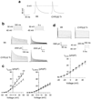
Fig. 1
Fig. 1
Characterization of action potentials and outward K+ currents in the CYP2J2 Tr and Wt cardiomyocytes. a, representative action potentials recorded from ventricular myocytes isolated from Wt and CYP2J2 Tr hearts. The action potentials were elicited by intracellular injection of depolarizing current pulses (15 pA with 10 ms duration) from a holding membrane potential of approximately −75 mV. b, superimposed original traces of transient outward K+ currents recorded from two representative cardiomyocytes isolated from Wt and CYP2J2 Tr mice. Whole-cell outward K+ currents were evoked by 300 ms (left) or 5 s (right) depolarizing pulses from −50 to +50 mV with 10-mV increments every 10 s. The membrane holding potential was set at −60 mV. c, current-voltage relationship of outward K+ currents recorded from single ventricular myocytes. Left, the peak amplitude of transient outward K+ currents (Ito,peak) was measured and averaged from individual cardiomyocytes (○, Wt, n 22; ●, CYP2J2 Tr, n = 23). Right, the late portion of transient outward K+ currents was measured at the point of 280 ms (Ito,280ms) and averaged from individual cardiomyocytes (○, Wt, n = 22; ●, CYP2J2 Tr, n = 23). The current density was calculated by the amplitude of current divided by the membrane capacitance of each cell. *, p < 0.05 versus Wt; **, p < 0.01 versus Wt. d, slowly inactivating outward K+ currents (IK,slow) recorded from isolated single ventricular myocytes. Top, current traces from representative Wt and CYP2J2 Tr cells elicited by test pulses from −40 to 50 mV with 10-mV increments every 10 s. A prepulse of 200 ms from the holding potential of −60 to +40 mV was applied and followed a 5-ms recovery interval to −60 mV. The protocol minimized contamination of transient outward current (Ito), the fast inactivating component of the depolarization-activated K+ currents. Bottom, the current-voltage relationships of IK,slow for Wt (○, n = 18) and CYP2J2 Tr (● n = 24) cardiomyocytes.
Fig. 2
Fig. 2
Comparison of voltage-gated Na+ currents in cardiomyocytes isolated from Wt and CYP2J2 Tr mice. a and b are superimposed whole-cell current traces recorded from single left ventricular cardiomyocytes isolated from Wt (a) and CYP2J2 Tr (b) mouse hearts. The currents were elicited by voltage pulses (10 ms duration every 5 s) from a holding potential of −80 mV down to −90 mV and up to 30 mV with 10-mV increments. c, current-voltage relationship curves for Wt (○ n = 15) and CYP2J2 Tr (●, n = 11) cardiomyocytes are shown. d, normalized steady-state inactivation of cardiac Na+ currents. Currents were elicited by a double-pulse protocol (inset) composed of a 10-ms test pulse to −30 mV after a 500 ms conditioning prepulse varying from −140 to −40 mV with 10-mV increments every 10 s from a holding potential of −80 mV. Peak Na+ currents elicited by test pulses were normalized to the maximal currents recorded with the prepulses from −140 to −40 mV and plotted against the prepulse voltages for the Wt (○) and CYP2J2 Tr (●) cardiomyocytes. The inactivation data points of peak INa were fitted to a Boltzmann equation.
Fig. 3
Fig. 3
Suppression of Ito,peak by the P450 epoxygenase inhibitor MS-PPOH in CYP2J2 Tr cardiomyocytes. Whole-cell outward K+ currents were evoked by 5-s (right panels) depolarizing pulses from −50 mV to 70 mV in 10 mV increments. The membrane holding potential was −60 mV, and the pulse rate was 0.1 Hz. The early portions (600 ms) of the currents are shown in the corresponding left panels (see the protocols in Fig. 1b). a, the effect of extracellular application of 25 µM MS-PPOH on outward K+ currents in a Wt cardiomyocyte. Addition of 2 mM 8-Br-cAMP after MS-PPOH application did not significantly alter the outward K+ currents. b, bath perfusion with 25 µM MS-PPOH significantly inhibited Ito,peak but did not affect the late portion of Ito (Ito,280ms) or the delayed outward K+ current (IK,slow) in a CYP2J2 Tr cardiomyocyte. Addition of 2 mM 8-Br-cAMP reversed the MS-PPOH-induced inhibition of Ito,peak. c, averaged data from multiple experiments showing inhibition of Ito,peak by 25 µM MS-PPOH in CYP2J2 Tr cardiomyocytes and reversal of this effect after washout. Currents were evoked by 5-s single-step pulses from −60 to 70 mV. The pulse rate was 0.1 Hz with a holding potential of −60 mV. The maximal peak amplitude of Ito was measured. Data are presented as mean ± S.E. ** p < 0.01 versus Wt.
Fig. 4
Fig. 4
Suppression of Ito,peak by an inhibitory CYP2J2 monoclonal antibody in CYP2J2 Tr cardiomyocytes. Whole-cell outward K+ currents were evoked by 5-s (right) depolarizing pulses from −50 to +70 mV in 10-mV increments. The membrane holding potential was −60 mV, and pulse rate was 0.1 Hz. The early portions (600 ms) of the currents are shown in the corresponding left panels (see the protocols in Fig. 1b). a, the effect of intracellular dialysis with MAb-1 (0.125 mg/ml IgG) on the outward K+ currents in a Wt cardiomyocyte. Addition of 40 nM 11,12-EET to the bath solution after MAb-1 dialysis did not significantly alter the outward K+ currents. b, intracellular dialysis with MAb-1 (0.125 mg/ml IgG) significantly inhibited Ito,peak but did not affect the late portion of Ito (Ito,280ms) or the delayed outward K+ current (IK,slow) in a CYP2J2 Tr cardiomyocyte. Addition of 40 nM 11,12-EET to the bath solution reversed the MAb-1-induced inhibition of Ito,peak. c, averaged data from multiple experiments showing that inhibition of Ito,peak by MAb-1 is significantly greater in CYP2J2 Tr cardiomyocytes than in Wt cardiomyocytes. Currents were evoked by 5-s single-step pulses from −60 to +70 mV. The pulse rate was 0.1 Hz with a holding potential of −60 mV. The maximal peak amplitude of Ito was measured. Data are presented as mean ± S.E. *, p < 0.05 versus Wt.
Fig. 5
Fig. 5
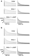
Suppression of Ito,peak by an inhibitory CYP2J2 monoclonal antibody in CYP2J2 Tr cardiomyocytes. Whole-cell outward K+ currents were evoked by 5 s (right) depolarizing pulses from −50 to 70 mV in 10-mV increments. The membrane holding potential was −60 mV and pulse rate was 0.1 Hz. The early portions (600 ms) of the currents are shown in the corresponding left panels (see the protocols in Fig. 1b). a, the effect of intracellular dialysis with MAb-1 (0.125 mg/ml IgG) on the outward K+ currents in a Wt cardiomyocyte. Addition of 2 mM 8-Br-cAMP to the bath solution after MAb-1 dialysis did not significantly alter the outward K+ currents. b, intracellular dialysis with the MAb-1 (0.125 mg/ml IgG) significantly inhibited the peak Ito (Ito,peak) but did not affect the late portion of Ito (Ito,280ms) or the delayed outward K+ current (IK,slow) in a CYP2J2 Tr cardiomyocyte. Addition of 2 mM 8-Br-cAMP to the bath solution reversed the MAb-1-induced inhibition of Ito,peak.
Fig. 6
Fig. 6
Effects of an inhibitory CYP2J2 monoclonal antibody on action potentials in Wt and CYP2J2 Tr cardiomyocytes. Top, representative action potentials recorded from a ventricular myocyte isolated from a Wt mouse heart. Intracellular dialysis with the MAb-1 (0.125 mg/ml IgG) slightly prolonged the duration of the action potential. Bottom, intracellular dialysis with MAb-1 (0.125 mg/ml IgG) markedly prolonged the duration of the action potential in a CYP2J2 Tr cardiomyocyte. Bath perfusion with 40 nM 11,12-EET almost completely reversed the MAb-1 effect. The action potentials were elicited by intracellular injection of depolarizing current pulses (15 pA with 10 ms duration) from a holding membrane potential of approximately −75 mV. Initial, the action potentials were recorded immediately after forming the whole-cell configuration. MAb-1, the action potentials were recorded 15 min after intracellular dialysis with MAb-1. EET, the action potential were recorded 10 min after bath perfusion with 11,12-EET.
Fig. 7
Fig. 7
Expression of voltage-gated K+ channels in CYP2J2 Tr and Wt hearts. a, lysates prepared from CYP2J2 Tr (n = 3) and Wt (n = 3) hearts were immunoblotted with selective antibodies to KChIP2, Kv1.4, Kv4.2, Kv4.3, and Na+/K+-ATPase α1 as described under Materials and Methods. Molecular masses are shown to the left of the panels. b, densitometry was performed and the expression of voltage-gated K+ channels was normalized to the expression of Na+/K+-ATPase α1.

Similar articles

Cited by

References

    1. Alvarez J, Montero M, Garcia-Sancho J. Cytochrome P450 may regulate plasma membrane Ca2+ permeability according to the filling state of the intracellular Ca2+ stores. FASEB J. 1992;6:786–792. - PubMed
    1. Anderson AE, Adams JP, Qian Y, Cook RG, Pfaffinger PJ, Sweatt JD. Kv4.2 phosphorylation by cyclic AMP-dependent protein kinase. J Biol Chem. 2000;275:5337–5346. - PubMed
    1. Brand-Schieber E, Falck JF, Schwartzman M. Selective inhibition of arachidonic acid epoxidation in vivo. J Physiol Pharmacol. 2000;51:655–672. - PubMed
    1. Campbell WB, Gebremedhin D, Pratt PF, Harder DR. Identification of epoxyeicosatrienoic acids as endothelium-derived hyperpolarizing factors. Circ Res. 1996;78:415–423. - PubMed
    1. Carroll MA, Doumad AB, Li J, Cheng MK, Falck JR, McGiff JC. Adenosine2A receptor vasodilation of rat preglomerular microvessels is mediated by EETs that activate the cAMP/PKA pathway. Am J Physiol Renal Physiol. 2006;291:F155–F161. - PubMed

Publication types

MeSH terms

Substances

LinkOut - more resources