. 2007;35(15):e95.
doi: 10.1093/nar/gkm540. Epub 2007 Jul 25.

Improved identification of enriched peptide RNA cross-links from ribonucleoprotein particles (RNPs) by mass spectrometry

Affiliations

Improved identification of enriched peptide RNA cross-links from ribonucleoprotein particles (RNPs) by mass spectrometry

Eva Kühn-Hölsken et al. Nucleic Acids Res. 2007.

Abstract

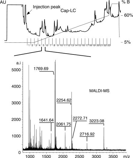
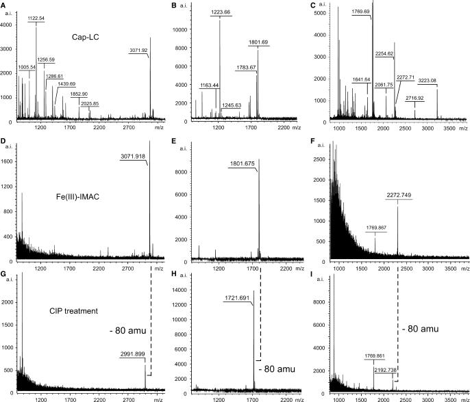
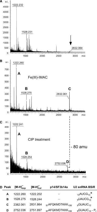
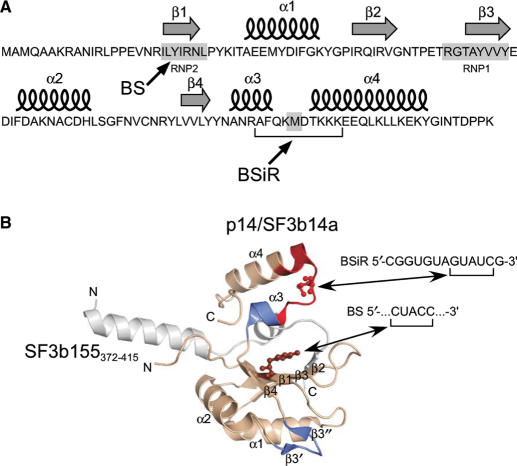
Direct UV cross-linking combined with mass spectrometry (MS) is a powerful tool to identify hitherto non-characterized protein-RNA contact sites in native ribonucleoprotein particles (RNPs) such as the spliceosome. Identification of contact sites after cross-linking is restricted by: (i) the relatively low cross-linking yield and (ii) the amount of starting material available for cross-linking studies. Therefore, the most critical step in such analyses is the extensive purification of the cross-linked peptide-RNA heteroconjugates from the excess of non-crosslinked material before MS analysis. Here, we describe a strategy that combines small-scale reversed-phase liquid chromatography (RP-HPLC) of UV-irradiated and hydrolyzed RNPs, immobilized metal-ion affinity chromatography (IMAC) to enrich cross-linked species and their analysis by matrix-assisted laser desorption/ionisation (MALDI) MS(/MS). In cases where no MS/MS analysis can be performed, treatment of the enriched fractions with alkaline phosphatase leads to unambiguous identification of the cross-linked species. We demonstrate the feasibility of this strategy by MS analysis of enriched peptide-RNA cross-links from UV-irradiated reconstituted [15.5K-61K-U4atac snRNA] snRNPs and native U1 snRNPs. Applying our approach to a partial complex of U2 snRNP allowed us to identify the contact site between the U2 snRNP-specific protein p14/SF3b14a and the branch-site interacting region (BSiR) of U2 snRNA.


Figures

Figure 1.
Figure 1.
Chromatogram of a capillary LC-run for the purification of peptide–RNA heteroconjugates derived from UV-irradiated U1 snRNP particles after hydrolysis with trypsin and RNase T1. The injection peak containing the moiety of non-crosslinked RNA oligonucleotides is indicated. A representative example of a MALDI-MS spectrum from a cap-LC fraction is shown.
Figure 2.
Figure 2.
Identification and validation of cross-linked peptide–RNA heteroconjugates by MALDI-ToF MS using IMAC and combined IMAC/CIP treatment. All spectra were recorded in positive ion-mode with DHB as matrix. (AC) MALDI-ToF MS spectra of cap-LC fractions derived from UV-irradiated [15.5K-61K-U4atac snRNA] complexes after hydrolysis with chymotrypsin and RNase T1 (A), chymotrypsin and RNases A/T1 (B) and from UV-irradiated native U1 snRNP particles after hydrolysis with trypsin and RNase T1 (C). (DF) MALDI-ToF MS spectra of the same fractions after treatment with IMAC. (GI) MALDI-ToF MS spectra of the same fractions after combined IMAC/CIP treatment. The masses of the enriched precursor ions at m/z 3071.918 and 1801.675 correspond to a chymotryptic fragment of the 61K (hPrp31) protein encompassing positions 263–273 (SSTSVLPHTGY) cross-linked to a CAUAG pentanucleotide of the U4atac snRNA (positions 42–46) (A,D,G) and to an AU dinucleotide of the U4atac snRNA (positions 43–44) (B,E,H), respectively, with and without the 3′-phosphate. The enriched precursor ion at m/z 2272.749 corresponds to a tryptic fragment of the U1 snRNP-specific 70K protein comprising positions 173–180 (RVLVDVER) cross-linked to an AUCA tetranucleotide of the U1 snRNA with and without the 3′-phosphate.
Figure 3.
Figure 3.
Identification and validation of a cross-linked peptide–RNA heteroconjugate derived from UV-irradiated [p14/SF3b14a-SF3b155282–424-U2 snRNA BSiR] complexes by MALDI-ToF MS using IMAC and combined IMAC/CIP treatment. All spectra were recorded in negative ion-mode with DHB as matrix. (A) MALDI-ToF spectrum of a cap-LC fraction derived from UV-irradiated [p14/SF3b14a-SF3b155282–424-U2 snRNA BSiR] complexes after hydrolysis with trypsin and RNase T1. The arrow indicates the signal of the cross-linked species (see panel B). (B) MALDI-ToF MS spectrum of the same fraction after treatment with IMAC. (C) MALDI-ToF MS spectrum of the same fraction after combined IMAC/CIP treatment. The masses of the enriched precursor ions (marked ‘C’ and ‘D’, respectively) correspond to a tryptic fragment of the p14/SF3b14a protein encompassing positions 97–106 (AFQKMDTKKK) cross-linked to a GUAUC pentanucleotide of the BSiR of U2 snRNA (positions 8–12) with and without the 3′-phosphate group. Ions marked ‘A’ and ‘B’ in panels B and C correspond to U2 snRNA BSiR oligonucleotides comprising positions 10–13 (AUCG) and 9–13 (UAUCG). Note that these oligonucleotides are located at the very 3′-end of the synthetic RNA oligomer and thus do not have a 3′-phosphate group accessible to CIP. (D) Assignment of the enriched precursor masses to peptide and RNA sequences. Asterisks indicate confirmation of RNA sequences by post-source decay (PSD) analysis.
Figure 4.
Figure 4.
Cross-linking sites in the 3D structure of p14/SF3b14a in complex with SF3b155373–415 according to ref. (39). (A) Secondary structure elements of p14/SF3b14a. Arrows indicate the amino acids cross-linked to the branch site [BS, (39)] and to the U2 branch-site interacting region (BSiR, this work). (B) Ribbon diagram of p14/SF3b14a in complex with SF3b155373–415. p14/SF3b14a is shown in beige, with Y22 in RNP2 (β1) that was cross-linked to the BS according to ref. (39) highlighted in balls-and-sticks, SF3b155373–415 is coloured grey. The peptide sequence (97AFQKMDTKKKEEQLK111) found to be cross-linked to the BSiR of U2 snRNA (this work) is marked in red with the putatively cross-linked amino acid M101 highlighted in balls-and-sticks. Regions marked in blue (β3′, β3″ and α3) were affected upon incubation with U2 BS/BSiR RNA duplex in NMR chemical shift experiments (31).

References

    1. Zhang B, Pan X, Cobb GP, Anderson TA. microRNAs as oncogenes and tumor suppressors. Dev. Biol. 2007;302:1–12. - PubMed
    1. Blencowe BJ. Alternative splicing: new insights from global analyses. Cell. 2006;126:37–47. - PubMed
    1. Jones-Rhoades MW, Bartel DP, Bartel B. MicroRNAs and their regulatory roles in plants. Annu. Rev. Plant Biol. 2006;57:19–53. - PubMed
    1. Kloosterman WP, Plasterk RH. The diverse functions of microRNAs in animal development and disease. Dev. Cell. 2006;11:441–450. - PubMed
    1. Mallory AC, Vaucheret H. Functions of microRNAs and related small RNAs in plants. Nat. Genet. 2006;38:S31–S36. Erratum in: Nat. Genet., 38, 850. - PubMed

Publication types

MeSH terms

Substances