. 2007 Nov;81(22):12535-42.
doi: 10.1128/JVI.00197-07. Epub 2007 Jul 25.

A recombinant Sendai virus is controlled by CD4+ effector T cells responding to a secreted human immunodeficiency virus type 1 envelope glycoprotein

Affiliations

A recombinant Sendai virus is controlled by CD4+ effector T cells responding to a secreted human immunodeficiency virus type 1 envelope glycoprotein

Scott A Brown et al. J Virol. 2007 Nov.

Abstract

The importance of antigen-specific CD4(+) helper T cells in virus infections is well recognized, but their possible role as direct mediators of virus clearance is less well characterized. Here we describe a recombinant Sendai virus strategy for probing the effector role(s) of CD4(+) T cells. Mice were vaccinated with DNA and vaccinia virus recombinant vectors encoding a secreted human immunodeficiency virus type 1 (HIV-1) envelope protein and then challenged with a Sendai virus carrying a homologous HIV-1 envelope gene. The primed mice showed (i) prompt homing of numerous envelope-primed CD4(+) T cell populations to the virus-infected lung, (ii) substantial production of gamma interferon, and interleukin-2 (IL-2), IL-4, and IL-5 in that site, and (iii) significantly reduced pulmonary viral load. The challenge experiments were repeated with immunoglobulin(-/-) microMT mice in the presence or absence of CD8(+) and/or CD4(+) T cells. These selectively immunodeficient mice were protected by primed CD4(+) T cells in the absence of antibody or CD8(+) T cells. Together, these results highlight the role of CD4(+) T cells as direct effectors in vivo and, because this protocol gives such a potent response, identify an outstanding experimental model for further dissecting CD4(+) T-cell-mediated immunity in the lung.


Figures

FIG. 1.
FIG. 1.
Immunodominant T-cell responses in HIV-1 envelope-immunized C57BL/6 mouse CD4+ T cells from C57BL/6 mice vaccinated with recombinant DNA (using a prime-boost regimen) were previously shown to respond predominantly toward the V3-C3 and C4 regions of the U92005 gp120 envelope protein (encompassed by peptide pools G and I, respectively [10] for peptide pool designations). Individual peptides within each positive pool were next tested using IFN-γ enzyme-linked immunospot assays with splenocytes enriched for CD4+ T cells from vaccinated and control animals. Positive responses are shown in this figure, as well as responses toward peptides flanking the positive peptide targets (data are represented as means ± standard error of the means).
FIG. 2.
FIG. 2.
Design and testing of the envelope recombinant Sendai virus. (A) The gp120 portion of the envelope gene was inserted between the P/C and M genes of the Sendai virus, as described in Materials and Methods. (B) A Western blot was prepared with purified wild-type Sendai virus (wt SeV, lane 1) or 5 μl of allantoic fluid from eggs infected with recombinant Sendai virus (rSeV-UG, lane 2). Samples were resolved by sodium dodecyl sulfate-polyacrylamide gel electrophoresis and transferred to nitrocellulose. Envelope protein was visualized by incubation with HIV-Ig (heat-inactivated HIV-infected human serum), followed by alkaline phosphatase-conjugated anti-human Ig antibody and a color reaction. gp140 protein from a lysate of transformed recombinant Chinese hamster ovary cells was applied to lane 3 (CHO gp140) as a positive control. Standard molecular masses (in kilodaltons) are indicated.
FIG. 3.
FIG. 3.
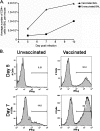
CD4+ T cells home to the site of recombinant Sendai virus challenge in vaccinated animals. (A) BAL fluid from vaccinated (DNA-vaccinia virus prime-boost regimen) and unvaccinated animals were collected on the indicated day following virus challenge (x axis), and cells were isolated. Pooled cells for each group were stained using anti-CD4 antibodies. The average total numbers of BAL fluid CD4+ lymphocytes per mouse are indicated (y axis, four to five animals/group). (B) BAL fluid cells were collected from naïve mice (left panels) and vaccinated mice (right panels) on day 5 (upper panels) and day 7 (lower panels) after recombinant Sendai virus challenge. Pooled BAL fluid cells were tested for responses to immunodominant peptides (IVGNIRQAHCNVSKA and GKAMYAPPIAGLIQC; 10 μg/ml per peptide) by intracellular cytokine staining following 5 h of restimulation in the presence of brefeldin A. Cells were gated on CD4+ T cells prior to flow cytometry analyses. A marker was set to indicate positive responses. Negative control responses among cells that were not stimulated with peptide in vitro were 0.76% and 1.76% for day 5 unvaccinated and vaccinated mice, respectively, and 12.6% and 1.01% for day 7 unvaccinated and vaccinated mice, respectively.
FIG. 4.
FIG. 4.
Both TH1 and TH2 cytokines were detected in the lungs of challenged animals. BAL fluid from mice vaccinated with a DNA-vaccinia virus prime-boost regimen and challenged with the Sendai virus recombinant were examined for IL-2, IFN-γ, IL-4, IL-5, and IL-10 content (on days 3, 5, 7, and 10 following challenge). Unvaccinated, challenged mice were used as controls. Each symbol represents an average response among animals (three to five animals were tested per group, and data are represented as concentration means ± standard error of the means). *, P < 0.05 using unpaired Student's t test or one sample t test.
FIG. 5.
FIG. 5.
Vaccinated animals control virus challenge. Vaccinated (DNA-vaccinia virus prime-boost regimen) and unvaccinated mice were challenged with recombinant Sendai virus (1 × 10⁶ PFU, i.n. administration). On each of days 3, 5, and 7 (x axis), groups of vaccinated and unvaccinated mice were sacrificed (four to five mice per group). Lungs were harvested and challenge virus titers were measured by plaque formation with LLC-MK2 cells, and data are represented as PFU means ± standard error of the means (PFU, y axis). *, P < 0.05 using unpaired Student's t test.
FIG. 6.
FIG. 6.
Virus clearance in the absence of antibody. Vaccinated (DNA-vaccinia virus prime-boost regimen) and unvaccinated μMT animals were challenged i.n. with 1 × 10⁵ PFU recombinant Sendai virus. Lungs were harvested on day 5 following challenge, and the titers of challenge virus in the lungs were determined by a TCID50 measurement with LLC-MK2 cells. The Reed-Muench formula was used to calculate the TCID50 value. Each symbol represents the TCID50 value of a different animal. *, P < 0.05 using unpaired Student's t test.
FIG. 7.
FIG. 7.
Depletion of CD4+ T cells in vaccinated μMT mice eliminates their ability to clear recombinant Sendai virus. μMT mice vaccinated with the DNA-vaccinia virus prime-boost regimen were treated with GK1.5 antibody (to remove CD4+ cells, ΔCD4) and/or 2.43 antibody (to remove CD8+ T cells, ΔCD8) on days −5, −3, −1, +1, and + 3 relative to challenge (see Materials and Methods for details). On day 5 following Sendai virus challenge, lungs were harvested to measure virus infection (by TCID50 measurements with LLC-MK2 cells). The Reed-Muench formula was used to calculate the TCID50 value. Each symbol represents the TCID50 value of a different animal. *, P < 0.05 using unpaired Student's t test.

References

    1. Abel, K., D. M. Rocke, B. Chohan, L. Fritts, and C. J. Miller. 2005. Temporal and anatomic relationship between virus replication and cytokine gene expression after vaginal simian immunodeficiency virus infection. J. Virol. 79:12164-12172. - PMC - PubMed
    1. Adhikary, D., U. Behrends, A. Moosmann, K. Witter, G. W. Bornkamm, and J. Mautner. 2006. Control of Epstein-Barr virus infection in vitro by T helper cells specific for virion glycoproteins. J. Exp. Med. 203:995-1006. - PMC - PubMed
    1. Allan, W., Z. Tabi, A. Cleary, and P. C. Doherty. 1990. Cellular events in the lymph node and lung of mice with influenza. Consequences of depleting CD4+ T cells. J. Immunol. 144:3980-3986. - PubMed
    1. Andreansky, S., H. Liu, H. Adler, U. H. Koszinowski, S. Efstathiou, and P. C. Doherty. 2004. The limits of protection by “memory” T cells in Ig-/- mice persistently infected with a gamma-herpesvirus. Proc. Natl. Acad. Sci. USA 101:2017-2022. - PMC - PubMed
    1. Belyakov, I. M., M. A. Derby, J. D. Ahlers, B. L. Kelsall, P. Earl, B. Moss, W. Strober, and J. A. Berzofsky. 1998. Mucosal immunization with HIV-1 peptide vaccine induces mucosal and systemic cytotoxic T lymphocytes and protective immunity in mice against intrarectal recombinant HIV-vaccinia challenge. Proc. Natl. Acad. Sci. USA 95:1709-1714. - PMC - PubMed

Publication types

MeSH terms