. 2007 Oct;81(19):10567-74.
doi: 10.1128/JVI.01181-07. Epub 2007 Jul 25.

Molecular determinants of antiviral potency of paramyxovirus entry inhibitors

Affiliations

Molecular determinants of antiviral potency of paramyxovirus entry inhibitors

M Porotto et al. J Virol. 2007 Oct.

Abstract

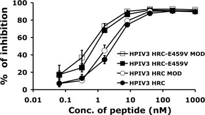
Hendra virus (HeV) and Nipah virus (NiV) constitute the Henipavirus genus of paramyxoviruses, both fatal in humans and with the potential for subversion as agents of bioterrorism. Binding of the HeV/NiV attachment protein (G) to its receptor triggers a series of conformational changes in the fusion protein (F), ultimately leading to formation of a postfusion six-helix bundle (6HB) structure and fusion of the viral and cellular membranes. The ectodomain of paramyxovirus F proteins contains two conserved heptad repeat regions, the first (the N-terminal heptad repeat [HRN]) adjacent to the fusion peptide and the second (the C-terminal heptad repeat [HRC]) immediately preceding the transmembrane domain. Peptides derived from the HRN and HRC regions of F are proposed to inhibit fusion by preventing activated F molecules from forming the 6HB structure that is required for fusion. We previously reported that a human parainfluenza virus 3 (HPIV3) F peptide effectively inhibits infection mediated by the HeV glycoproteins in pseudotyped-HeV entry assays more effectively than the comparable HeV-derived peptide, and we now show that this peptide inhibits live-HeV and -NiV infection. HPIV3 F peptides were also effective in inhibiting HeV pseudotype virus entry in a new assay that mimics multicycle replication. This anti-HeV/NiV efficacy can be correlated with the greater potential of the HPIV3 C peptide to interact with the HeV F N peptide coiled-coil trimer, as evaluated by thermal unfolding experiments. Furthermore, replacement of a buried glutamic acid (glutamic acid 459) in the C peptide with valine enhances antiviral potency and stabilizes the 6HB conformation. Our results strongly suggest that conserved interhelical packing interactions in the F protein fusion core are important determinants of C peptide inhibitory activity and offer a strategy for the development of more-potent analogs of F peptide inhibitors.


Figures

FIG. 1.
FIG. 1.
Inhibition of HeV and NiV infection by HPIV3 HRC peptides. Vero cell monolayers were infected with 50 to 80 PFU of HeV or NiV in the presence of HPIV wt peptides or HPIV scrambled (sc) peptides at the indicated peptide concentrations (nM). Values are means (±standard deviations; n = 6).
FIG. 2.
FIG. 2.
Inhibition of HeV and NiV plaque enlargement by HPIV3-derived and NiV-derived peptides. Vero cell monolayers were infected with 50 to 80 PFU of HeV or NiV. At 30 min after infection, HPIV wt peptides, HPIV scrambled (sc) peptides, or NiV-derived peptides (NiV FC2) were added at the indicated concentrations (nM). Plaque size (pixel2) is also shown. Values are means (±standard deviations; n = 6).
FIG. 3.
FIG. 3.
Inhibitory effects of HRC peptides (wt HPIV3 and the HPIV3 E459V mutated peptide) on infection by HeV G/F pseudotyped viruses. Vero cells were infected with pseudotyped VSV-ΔG-RFP-HeV G-CT32/F virus at an MOI of 0.25 in the presence of increasing concentrations (Conc.) of peptide inhibitors. At 36 h after infection, the number of fluorescent cells was determined using FACS analysis. The results are shown as percent inhibition of viral entry (compared to results for inhibitor-free controls). Values are means (±standard deviations) of results from three separate experiments.
FIG. 4.
FIG. 4.
Inhibition by HPIV3 HRC peptides of infection with HPIV3 virions. CV-1 cells were infected with HPIV3 wt virus at an MOI of 6 × 10⁻⁴ in the presence and absence of different concentrations (Conc.) of peptides. After 2 h, cells were overlaid with agarose, and plaques were stained at 18 h postinfection. The percent inhibition of viral entry (compared to results for control cells infected in the absence of inhibitors) is shown as a function of the concentration (log scale) of HPIV3 HRC peptide with and without Cap or mutated at position 459 with or without Cap. Data points are means (±standard deviations) of results from three separate experiments. MOD, modified.
FIG. 5.
FIG. 5.
Inhibition of HeV pseudotyped-virus infection by modified HPIV3 F HRC peptides, determined by use of an assay that mimics multicycle replication. Vero cells transfected with HeV G and HeV F were infected with pseudotyped VSV-ΔG-RFP-HeV G-CT32/F virus in the presence of HPIV3 HRC and HPIV3 E459V HRC at various concentrations (Conc.), either in modified (N-acetylated and capped) (MOD) or in unmodified form. At 36 h after infection, pseudotyped viral entry was quantified by assessing red fluorescence through FACS analysis. Values are means (±standard deviations) of results from three separate experiments.
FIG. 6.
FIG. 6.
Interactions of two hydrophobic HR (HRN and HRC) regions in HeV and HPIV3 F. (A) Amino acid sequences of the N42 and C33 segments of the homotypic and chimeric HRN/HRC constructs. The recombinant N42(L6)C33 peptide consists of the N44 and C33 segments connected by the linker residues Ser-Gly-Gly-Ser-Gly-Gly. A glutamic acid 459-to-valine mutation is indicated in the E459V sequence. (B) Thermal melts of N42HeV(L6)C33HeV (triangles), N42HeV(L6)C33HPIV3 (circles), and E459V (squares) constructs, monitored by the CD signal at 222 nm at a 50 μM protein concentration in TBS (pH 8.0) in the presence of 3 M GuHCl, a chemical denaturant. deg, degrees. (C) Sedimentation equilibrium data (19,000 rpm) for the E459V peptide, collected at 20°C in TBS (pH 8.0) at an ∼200 μM protein concentration. The deviation in the data from the linear fit for a trimeric model is plotted (top).

Similar articles

Cited by

References

    1. Aguilar, H. C., K. A. Matreyek, C. M. Filone, S. T. Hashimi, E. L. Levroney, O. A. Negrete, A. Bertolotti-Ciarlet, D. Y. Choi, I. McHardy, J. A. Fulcher, S. V. Su, M. C. Wolf, L. Kohatsu, L. G. Baum, and B. Lee. 2006. N-glycans on Nipah virus fusion protein protect against neutralization but reduce membrane fusion and viral entry. J. Virol. 80:4878-4889. - PMC - PubMed
    1. Anfinsen, C. B. 1973. Principles that govern the folding of protein chains. Science 181:223-230. - PubMed
    1. Arakawa, T., and S. N. Timasheff. 1984. Protein stabilization and destabilization by guanidinium salts. Biochemistry 23:5924-5929. - PubMed
    1. Baker, K. A., R. E. Dutch, R. A. Lamb, and T. S. Jardetzky. 1999. Structural basis for paramyxovirus-mediated membrane fusion. Mol. Cell 3:309-319. - PubMed
    1. Bossart, K. N., B. A. Mungall, G. Crameri, L. F. Wang, B. T. Eaton, and C. C. Broder. 2005. Inhibition of Henipavirus fusion and infection by heptad-derived peptides of the Nipah virus fusion glycoprotein. Virol. J. 2:57. - PMC - PubMed

Publication types

MeSH terms

LinkOut - more resources