. 2007 Oct;81(19):10804-14.
doi: 10.1128/JVI.00999-07. Epub 2007 Jul 25.

Single amino acid changes in the Nipah and Hendra virus attachment glycoproteins distinguish ephrinB2 from ephrinB3 usage

Affiliations

Single amino acid changes in the Nipah and Hendra virus attachment glycoproteins distinguish ephrinB2 from ephrinB3 usage

Oscar A Negrete et al. J Virol. 2007 Oct.

Abstract

The henipaviruses, Nipah virus (NiV) and Hendra virus (HeV), are lethal emerging paramyxoviruses. EphrinB2 and ephrinB3 have been identified as receptors for henipavirus entry. NiV and HeV share similar cellular tropisms and likely use an identical receptor set, although a quantitative comparison of receptor usage by NiV and HeV has not been reported. Here we show that (i) soluble NiV attachment protein G (sNiV-G) bound to cell surface-expressed ephrinB3 with a 30-fold higher affinity than that of sHeV-G, (ii) NiV envelope pseudotyped reporter virus (NiVpp) entered ephrinB3-expressing cells much more efficiently than did HeV pseudotyped particles (HeVpp), and (iii) NiVpp but not HeVpp entry was inhibited efficiently by soluble ephrinB3. These data underscore the finding that NiV uses ephrinB3 more efficiently than does HeV. Henipavirus G chimeric protein analysis implicated residue 507 in the G ectodomain in efficient ephrinB3 usage. Curiously, alternative versions of published HeV-G sequences show variations at residue 507 that can clearly affect ephrinB3 but not ephrinB2 usage. We further defined surrounding mutations (W504A and E505A) that diminished ephrinB3-dependent binding and viral entry without compromising ephrinB2 receptor usage and another mutation (E533Q) that abrogated both ephrinB2 and -B3 usage. Our results suggest that ephrinB2 and -B3 binding determinants on henipavirus G are distinct and dissociable. Global expression analysis showed that ephrinB3, but not ephrinB2, is expressed in the brain stem. Thus, ephrinB3-mediated viral entry and pathology may underlie the severe brain stem neuronal dysfunction seen in fatal Nipah viral encephalitis. Characterizing the determinants of ephrinB2 versus -B3 usage will further our understanding of henipavirus pathogenesis.


Figures

FIG. 1.
FIG. 1.
Construction, expression, and receptor binding analysis of sNiV-G and sHeV-G. (A) NiV-G and HeV-G were codon optimized and synthesized as described in Materials and Methods. The ectodomain of NiV-G (amino acid residues 71 to 602) was C-terminally tagged with an HA tag and placed under the control of a CMV promoter (sNiV-G). A similar construct was made using the HeV-G ectodomain (amino acid residues 71 to 604) (sHeV-G). Since G is a type II transmembrane protein, the kappa light chain signal sequence was placed at the N termini of both sNiV-G and sHeV-G to promote secretion of soluble protein into the supernatant. A linker sequence was also added between the kappa leader and the start of the G ectodomain. (B) Various oligomeric forms that likely correspond to monomeric (M), dimeric (D), and tetrameric (T) species of sG-HA were seen after the proteins were separated by reducing and nonreducing SDS-PAGE and visualized by anti-HA Western blot analysis. (C and D) EphrinB2 and -B3 stably transfected CHOpgsA745 cells (CHO-B2 and CHO-B3 cells, respectively) were used to measure sNiV-G (filled squares) and sHeV-G (open triangles) cell surface binding. Increasing concentrations of sG-HA (10−12 to 10−7 M) were added to CHO-B2 or CHO-B3 cells, and binding was assessed by flow cytometry using anti-HA monoclonal antibodies and R-phycoerythrin-conjugated anti-mouse IgG. Regression curves were generated based on values displayed as percentages of maximal binding, with the saturation value set to 100%. Each data point is an average of triplicates ± standard deviation (SD).
FIG. 2.
FIG. 2.
HeV uses ephrinB3 less efficiently than does NiV. (A and B) Homotypic envelope-mediated infections were performed using NiV-G and NiV-F (NiVpp) or HeV-G and HeV-F (HeVpp) glycoproteins pseudotyped into a VSV-ΔG-Luc reporter virus. Heterotypic envelope-mediated infections were performed using NiV-G and HeV-F (NG/HF) or HeV-G and NiV-F (HG/NF) pseudotyped into the VSV reporter virus. Equivalent amounts of viral particles (titrated to give equivalent Luc activities on CHO-B2 cells) were used to infect parental CHOpgsA745 cells (CHO) or CHO-pgsA745 cells stably expressing human ephrinB1, -B2, and -B3 (CHO-B1, CHO-B2, and CHO-B3 cells, respectively). CHO-B2 and CHO-B3 cells expressed ephrinB2 and -B3, respectively, at similar levels (see Materials and Methods). Entry of homotypic and heterotypic viruses was measured by quantifying Renilla Luc activity according to the manufacturer's directions. Relative light units (RLU) were acquired and quantified on a luminometer. Data shown are averages for three independent experiments ± SD. (C and D) Dilutions of homotypic (NiVpp and HeVpp) and heterotypic (NG/HF and HG/NF) reporter viruses were made to span 2 log and subsequently used for viral entry into CHO-B2 and CHO-B3 cells. The amount of pseudotyped virus entry was measured in RLU. Data shown are averages for three independent experiments ± SD. Error bars are too small to be seen on the log scale presented for the RLU data.
FIG. 3.
FIG. 3.
Soluble ephrin-B3 inhibits HeVpp less efficiently than NiVpp. NiV-F/G (NiVpp) and HeV-F/G (HeVpp) VSV-ΔG-Luc pseudotyped viruses were used to infect CHO-B2 cells in the presence of increasing amounts of soluble ephrinB2-Fc (10−12 to 10−8 M) and ephrinB3-Fc (10−13 to 10−7 M) fusion proteins (B2-Fc and B3-Fc, respectively). Entry was measured as described in the legend to Fig. 2A. Inhibition curves were generated via GraphPad Prism. Data are the averages for three independent experiments ± SD.
FIG. 4.
FIG. 4.
Determinant of ephrinB3 usage maps to a single amino acid in the G protein. (A) Soluble chimeric G proteins were made by N-terminally fusing the indicated segments from the ectodomain of sNiV-G to the corresponding remaining C-terminal segment from sHeV-G. (B) Equal amounts of sHeV-G, sNiV-G, and the chimeric G proteins were incubated with CHO-B2 or CHO-B3 cells. The amount of cell surface binding was measured by flow cytometry and calculated as the MFI. (C) Full-length chimeric HeV-G proteins were made by replacing residues 494 to 573 (HN494-573), 512 to 573 (HN494-573), or 494 to 512 (HN494-512) with homologous NiV-G residues. (D) HeV-G, NiV-G, and the full-length chimeric G proteins were used to make a VSV-Luc reporter virus, which was used to infect CHO-B2 or CHO-B3 cells. The amount of entry was measured in RLU, based on the amount of Luc activity present in the cells 24 h after infection. (E) Single amino acid substitutions were made in full-length HeV-G at residues 498 (H-V498T), 502 (H-V502I), and 507 (H-S507V) to complement the NiV-G residues at the equivalent positions. (F) The H-V498T, H-V502I, and H-S507V mutants were used to make pseudotyped VSV, which was used, along with NiVpp and HeVpp, to infect CHO-B2 or CHO-B3 cells. All data points in panels B, D, and F are averages of triplicates ± SD.
FIG. 5.
FIG. 5.
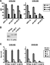
A mutant with a serine-to-threonine change at residue 507 in HeV-G gains ephrinB3 binding and viral entry abilities comparable to those of NiV. (A) sNiV-G and sHeV-G with residue T507 (sH-507T) or S507 (sH-507S), used at 2 nM, 0.4 nM, and 0.1 nM concentrations, were allowed to bind independently to CHO-B2 or CHO-B3 cells. The amount of binding was assessed by flow cytometry using anti-HA monoclonal antibodies and R-phycoerythrin-conjugated anti-mouse IgG. Data are the averages of triplicates ± SD. (B) Henipavirus envelope glycoproteins, namely, NiV-F/G (NiVpp) or HeV-F/G with residue 507T or 507S (H-507T or H-507S, respectively), were pseudotyped onto VSV reporter viruses. NiVpp, H-507T, and H-507S virus particles were run in a reducing SDS-PAGE gel and detected by anti-HA (αHA) Western blotting to detect G. Note that HeV-G always runs slightly higher on the gel than NiV-G. (C) Fivefold serial dilutions were made with NiVpp, H-507T, and H-507S VSV reporter viruses and subsequently used to infect CHO-B2 and CHO-B3 cells at similar concentrations, based on the amount of G incorporated onto the viral particles (B). Luc reporter activity was measured in RLU. Data are the averages for three independent experiments ± SD.
FIG. 6.
FIG. 6.
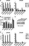
Residues near amino acid 507 in NiV-G are involved in distinct ephrinB3 binding. (A) Flow cytometry was used to assess the binding of cell surface-expressed ephrinB2 and -B3 with wild-type sNiV-G or sNiV-G with a mutation at W504A (sN-W504A), E505A (sN-E505A), V507S (sN-V507S), or E533Q (sN-E533Q). The amounts of binding of various concentrations (2 nM, 0.4 nM, and 0.1 nM) of wild-type or mutant sNiV-G to CHO-B2 and CHO-B3 cells are shown as MFI values. Data are the averages of triplicates ± SD. (B) Full-length wild-type NiV-G or mutant G proteins (W504A, E505A, V507S, and E533Q) were expressed in 293T cells and used for binding analysis with two nonconformational (anti-HA and 806) and two conformational (26 and 45) antibodies. All values were normalized to the MFI of the wild-type protein, set at 100% (data shown are averages of triplicates ± SD). (C) NiV envelope glycoproteins F and wild-type (WT) G (NiVpp) or NiV-F and mutant G (N-W504A, N-E505A, N-V507S, and N-E533Q) were pseudotyped onto VSV-ΔG-Luc reporter viruses. Wild-type NiV and mutant virus particles were run in a reducing SDS-PAGE gel and detected by anti-HA (αHA) Western blotting to detect G. (D) Dilutions (1×, 5×, and 25×) of wild-type NiV and mutant VSV reporter viruses were used to infect CHO-B2 and CHO-B3 cells at similar concentrations, based on the amount of G incorporated onto the viral particles (C). Data are the averages for three independent experiments ± SD.
FIG. 7.
FIG. 7.
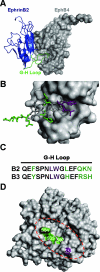
NiV-G model and proposed site of ephrinB2 and -B3 interaction in comparison to the crystal structure of the ephrinB2-EphB4 complex. (A) The crystal structure of an ephrinB2 monomer (blue cartoon representation) in complex with its cognate receptor, EphB4 (surface representation), indicates that the G-H loop of ephrinB2 (green) inserts into a hydrophobic canyon of EphB4 (11). (B) Detailed view of the G-H loop of ephrinB2 (stick representation) that makes contact with the binding cleft of EphB4 (surface representation). (C) Alignment of the G-H loop residues of ephrinB2 and -B3. Color-coded residues correspond to those indicated in panel B. The purple residues are critical for ephrinB2 and -B3 binding to NiV-G, and the green residues indicate the differences between ephrinB2 and -B3 in the G-H loop. (D) Structural model of the NiV-G protein globular domain described by Guillaume et al. (20), displayed as a top view with surface representation. Green residues (W504, E505, and V507) localize to the top surface of the NiV-G model and represent residues involved in distinct ephrinB3 binding. The purple residue (E533) has overlapping ephrinB2 and -B3 binding properties. The dashed red oval highlights a predicted binding cleft or canyon in the NiV-G model. All images were created using PyMol v0.99 (DeLano Scientific LLC, Palo Alto, CA).
FIG. 8.
FIG. 8.
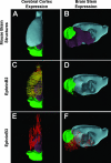
EphrinB2 and -B3 are coexpressed in the cerebral cortex, but ephrinB3 is distinctly expressed in the brain stem. (A and B) A three-dimensional model of the adult mouse brain was colored differently to represent the three major brain regions, i.e., the cerebral cortex (light blue), the cerebellum (green), and the brain stem (purple). The left hemisphere of the mouse brain was viewed from either a dorsal plane (A, C, and E) or a sagittal plane (B, D, and F). EphrinB2 expression in the cerebral cortex (C) or the brain stem (D) is indicated by orange and yellow dots. EphrinB3 expression in the cerebral cortex (E) or the brain stem (F) is displayed as red dots. All images were created using Brain Explorer v1.3, the Allen Brain Atlas (28) gene expression viewer program.

Similar articles

Cited by

References

    1. AbuBakar, S., L. Y. Chang, A. R. Ali, S. H. Sharifah, K. Yusoff, and Z. Zamrod. 2004. Isolation and molecular identification of Nipah virus from pigs. Emerg. Infect. Dis. 10:2228-2230. - PMC - PubMed
    1. Aguilar, H. C., O. A. Negrete, K. A. Matreyek, D. Y. Choi, and B. Lee. 2006. Annu. Meet. Am. Soc. Virol., abstr. P19-4. American Society for Virology, Madison, WI.
    1. Anonymous. 2004. Person-to-person transmission of Nipah virus during outbreak in Faridpur District, 2004. Health Sci. Bull. 2:5-9.
    1. Benson, M. D., M. I. Romero, M. E. Lush, Q. R. Lu, M. Henkemeyer, and L. F. Parada. 2005. Ephrin-B3 is a myelin-based inhibitor of neurite outgrowth. Proc. Natl. Acad. Sci. USA 102:10694-10699. - PMC - PubMed
    1. Bishop, K. A., T. S. Stantchev, A. C. Hickey, D. Khetawat, K. N. Bossart, V. Kerasnoperov, P. Gill, Y. R. Feng, L. Wang, B. Eaton, L. Wang, and C. C. Broder. 2007. Identification of Hendra virus G glycoprotein residues that are critical for receptor binding. J. Virol. 81:5893-5901. - PMC - PubMed

Publication types

MeSH terms

LinkOut - more resources