. 2007 Sep;17(9):1344-52.
doi: 10.1101/gr.6421207. Epub 2007 Jul 25.

Analysis of the VSG gene silent archive in Trypanosoma brucei reveals that mosaic gene expression is prominent in antigenic variation and is favored by archive substructure

Affiliations

Analysis of the VSG gene silent archive in Trypanosoma brucei reveals that mosaic gene expression is prominent in antigenic variation and is favored by archive substructure

Lucio Marcello et al. Genome Res. 2007 Sep.

Abstract

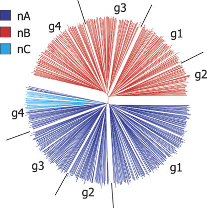
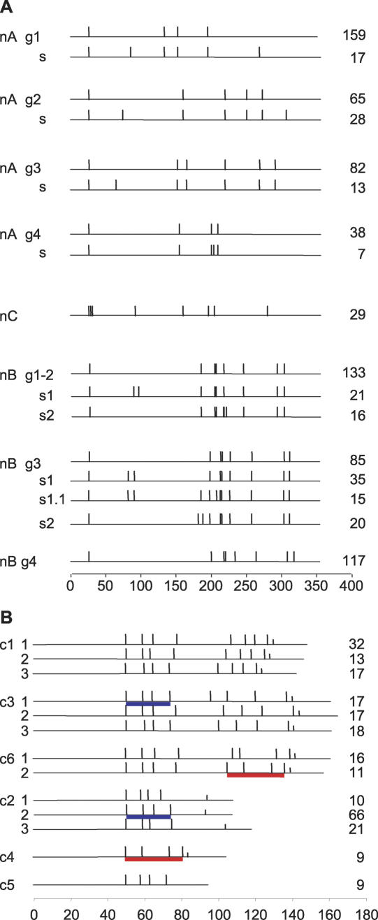
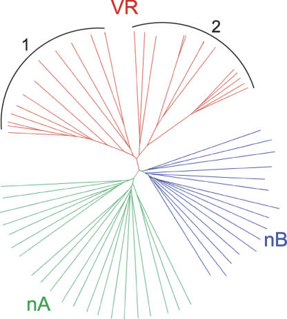
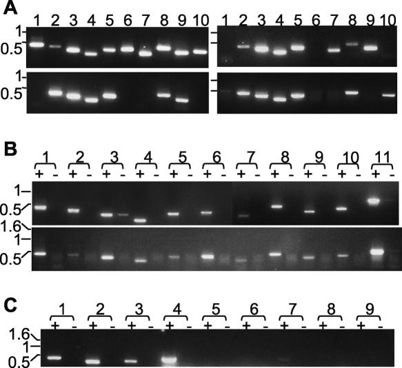
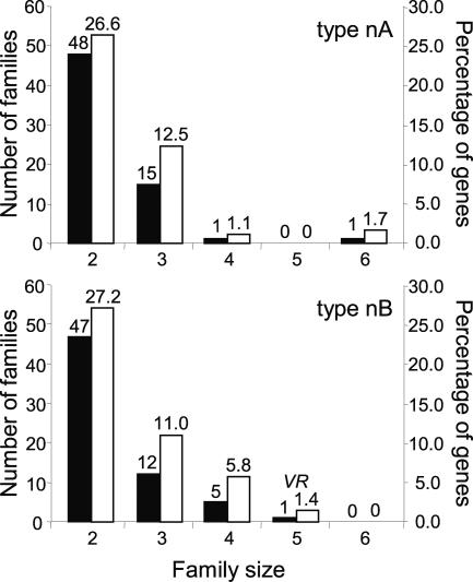
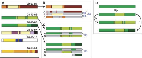
Trypanosoma brucei evades host acquired immunity through differential activation of its large archive of silent variant surface glycoprotein (VSG) genes, most of which are pseudogenes in subtelomeric arrays. We have analyzed 940 VSGs, representing one half to two thirds of the arrays. Sequence types A and B of the VSG N-terminal domains were confirmed, while type C was found to be a constituent of type A. Two new C-terminal domain types were found. Nearly all combinations of domain types occurred, with some bias to particular combinations. One-third of encoded N-terminal domains, but only 13% of C-terminal domains, are intact, indicating a particular need for silent VSGs to gain a functional C-terminal domain to be expressed. About 60% of VSGs are unique, the rest occurring in subfamilies of two to four close homologs (>50%-52% peptide identity). We found a subset of VSG-related genes, differing from VSGs in genomic environment and expression patterns, and predict they have distinct function. Almost all (92%) full-length array VSGs have the partially conserved flanks associated with the duplication mechanism that activates silent genes, and these sequences have also contributed to archive evolution, mediating most of the conversions of segments, containing >/=1 VSG, within and between arrays. During infection, intact array genes became activated by duplication after two weeks, and mosaic VSGs assembled from pseudogenes became expressed by week three and predominated by week four. The small subfamily structure of the archive appears to be fundamental in providing the interacting donors for mosaic formation.


Figures

Figure 1.
Figure 1.
Diversity of VSG N-terminal domain. Neighbor-joining tree based on ClustalX-generated multiple sequence alignment of predicted peptide sequences of 725 sequences. The three domain types are colored individually (by HyperTree) as shown in the key. g represents each group depicted in Fig. 2A.
Figure 2.
Figure 2.
Cysteine pattern of VSG domain types. Cys residue positions are shown as vertical bars, and the number of sequences in each category is shown to the right. Scale bars (number of residues) are shown for each panel. (A) N-terminal domain. g, group; s, subgroup. (B) Examples of C-terminal domain. Cys subdomains shared between different domain types are indicated by underlines of the same color. Two to three examples of domain type subgroups are given for the main domain types. Half-length vertical bars represent putative GPI signal sequences.
Figure 3.
Figure 3.
Tree of amino acid alignment of putative VSG-related proteins. The tree compares predicted products of 35 full-length, functional VSGs with those of 29 VR genes. The VR set falls into two clusters, denoted by 1 and 2.
Figure 4.
Figure 4.
Analysis of VR genes. (A) Presence of VR sequences in several T. brucei strains. PCR analysis of genomic DNA was undertaken with primers specific for: lanes 1–10, VR 1, 2, 4, 5, 8, 9, 11, 13, 15, and 18. T. brucei genomic DNA sources: top left, TREU 927; top right, Lister 427; bottom left, STIB 247; bottom right, EATRO 795. (B) Lack of stage-specificity of VR transcripts. RT-PCR analysis of VR transcripts represented in oligo[dT]-primed cDNA from bloodstream stage (top) and procyclic stage (bottom) TREU 927. Lane pairs 1–10: primers for VR 1, 2, 4, 5, 8, 9, 11, 13, 15, and 18; lane pairs 11: tubulin primers. In each lane pair, “+” denotes presence, and “−” denotes absence, of reverse transcriptase at the cDNA synthesis step. (C) Analysis of whether transcripts of alleles of VR and VSG genes are mutually exclusive. RT-PCR analysis of Lister 427 bloodstream trypanosome clone expressing linked HYG and 221 VSG genes, grown in hygromycin. oligo[dT]-primed cDNA was subjected to PCR using (lane pairs 1–9) primers for VR 2, VR 5, VR 15, 221 VSG, 118 VSG, VO2 VSG, 121 VSG, G4 VSG, S8 VSG. All images are of ethidium bromide-stained gels, and DNA size markers (kb) are indicated to the left of each.
Figure 5.
Figure 5.
Array evolution through duplication. For 31 VSG in the chr 8L array, which are depicted by boxes (followed by an orientation indicator) in the map at the top, the number of other copies of each in the genome (≥75% nucleotide identity) is shown by color intensity in the lower graphic, according to the tone key. The ends of duplicated sequences are aligned between the two graphics, demonstrating that these ends often coincide with the ends of VSGs.
Figure 6.
Figure 6.
VSG families. Pairwise comparison within N-terminal domain types. Peptide sequences encoded by 361 type nA VSGs were all scored against each other in ClustalW multiple sequence alignment, and 345 type nB VSGs were compared in the same way (bottom chart). The number of families (left scale of Y-axis) of given size (X-axis) are depicted in black, and the percentage of genes (right scale of Y-axis) within each domain type is depicted in white. VR indicates the family of VSG-related genes.
Figure 7.
Figure 7.
Structure and origin of putative mosaic cDNAs. (A) Mosaic structure. The seven detected mosaics for which significant donor information is available. Colors indicate patches matching different donors. The white regions in mosaic 28-10-10 indicate where no donor has been identified. (B) Origin and structure of N-terminal domain mosaic 22-07-02 from mouse 7 on day 22. The three putative silent donor genes (A, B, C) are shown below the cDNA, as thinner rectangles. Colors represent patches in common between donor and cDNA (positions of junctions in cDNA are averages where donors are identical in that region). The percentage nucleotide identity between silent donors is shown to the right. Vertical red bars depict frameshifts or stop codons in donor genes. The likely donors are: A, Tb11.v4.0074; B, Tb10.v4.0161; C, Tb11.14.0001. (C) Origin and structure of related mosaics coexisting in mouse 10 on day 28. Diagram is constructed as in B. For both mosaics, likely donors A and B are Tb11.09.0005 and Tb11.13.0003. For 28-10-03, likely donor C is read AZ217061. (D) Deduced pathway for development of related mosaics in mouse 10 on day 28.

References

    1. Aline R., Macdonald G., Brown E., Allison J., Myler P., Rothwell V., Stuart K., Macdonald G., Brown E., Allison J., Myler P., Rothwell V., Stuart K., Brown E., Allison J., Myler P., Rothwell V., Stuart K., Allison J., Myler P., Rothwell V., Stuart K., Myler P., Rothwell V., Stuart K., Rothwell V., Stuart K., Stuart K. (TAA)n within sequences flanking several intrachromosomal variant surface glycoprotein genes in Trypanosoma brucei. Nucleic Acids Res. 1985;13:3161–3177. - PMC - PubMed
    1. Aury J.M., Jaillon O., Duret L., Noel B., Jubin C., Porcel B.M., Segurens B., Daubin V., Anthouard V., Aiach N., Jaillon O., Duret L., Noel B., Jubin C., Porcel B.M., Segurens B., Daubin V., Anthouard V., Aiach N., Duret L., Noel B., Jubin C., Porcel B.M., Segurens B., Daubin V., Anthouard V., Aiach N., Noel B., Jubin C., Porcel B.M., Segurens B., Daubin V., Anthouard V., Aiach N., Jubin C., Porcel B.M., Segurens B., Daubin V., Anthouard V., Aiach N., Porcel B.M., Segurens B., Daubin V., Anthouard V., Aiach N., Segurens B., Daubin V., Anthouard V., Aiach N., Daubin V., Anthouard V., Aiach N., Anthouard V., Aiach N., Aiach N., et al. Global trends of whole-genome duplications revealed by the ciliate Paramecium tetraurelia. Nature. 2006;444:171–178. - PubMed
    1. Barbet A.F., Kamper S.M., Kamper S.M. The importance of mosaic genes to trypanosome survival. Parasitol. Today. 1993;9:63–66. - PubMed
    1. Barry J.D., McCulloch R., McCulloch R. Antigenic variation in trypanosomes: Enhanced phenotypic variation in a eukaryotic parasite. Adv. Parasitol. 2001;49:1–70. - PubMed
    1. Barry J.D., Ginger M.L., Burton P., McCulloch R., Ginger M.L., Burton P., McCulloch R., Burton P., McCulloch R., McCulloch R. Why are parasite contingency genes often associated with telomeres? Int. J. Parasitol. 2003;33:29–45. - PubMed

Publication types

MeSH terms

Substances

LinkOut - more resources