. 2007 Oct;18(10):3873-82.
doi: 10.1091/mbc.e07-03-0205. Epub 2007 Jul 25.

Role of the V-ATPase in regulation of the vacuolar fission-fusion equilibrium

Affiliations

Role of the V-ATPase in regulation of the vacuolar fission-fusion equilibrium

Tonie L Baars et al. Mol Biol Cell. 2007 Oct.

Abstract

Like numerous other eukaryotic organelles, the vacuole of the yeast Saccharomyces cerevisiae undergoes coordinated cycles of membrane fission and fusion in the course of the cell cycle and in adaptation to environmental conditions. Organelle fission and fusion processes must be balanced to ensure organelle integrity. Coordination of vacuole fission and fusion depends on the interactions of vacuolar SNARE proteins and the dynamin-like GTPase Vps1p. Here, we identify a novel factor that impinges on the fusion-fission equilibrium: the vacuolar H(+)-ATPase (V-ATPase) performs two distinct roles in vacuole fission and fusion. Fusion requires the physical presence of the membrane sector of the vacuolar H(+)-ATPase sector, but not its pump activity. Vacuole fission, in contrast, depends on proton translocation by the V-ATPase. Eliminating proton pumping by the V-ATPase either pharmacologically or by conditional or constitutive V-ATPase mutations blocked salt-induced vacuole fragmentation in vivo. In living cells, fission defects are epistatic to fusion defects. Therefore, mutants lacking the V-ATPase display large single vacuoles instead of multiple smaller vacuoles, the phenotype that is generally seen in mutants having defects only in vacuolar fusion. Its dual involvement in vacuole fission and fusion suggests the V-ATPase as a potential regulator of vacuolar morphology and membrane dynamics.


Figures
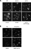
Figure 1.
Figure 1.
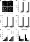
Vacuolar morphology of V-ATPase mutants. (A) vma1Δ, vma3Δ, vph1Δ, and WT (BY4741) yeast cells were grown in YPD, pH 5.5, to logarithmic phase, stained with FM4-64, and analyzed by spinning disk confocal microscopy by using an excitation laser at 488 nm and a 100× objective. As an alternative means to visualize vacuolar membranes, GFP-Pho8p was expressed in vph1Δ cells and revealed by fluorescence microscopy as described above. (B) Vacuolar membranes of wild-type and vph1Δcells (in BJ3505 and W303a background) were labeled with FM4-64 and visualized by fluorescence microscopy.
Figure 2.
Figure 2.
Fusion activity of V0 mutants. Vacuoles were prepared from WT and mutant cells (vma6Δ and vph1Δ). Their fusion activity was assayed in standard reactions run in the presence or absence of recombinant 0.1 g l−1 Vam7p. After 60 min on ice or at 27°C, alkaline phosphatase (ALP) activities were determined. ALP activity of the wild-type control at 27°C was 3–5 U. Ice values varied from 0.2 to 0.35 U, and they were subtracted from the respective 27°C values.
Figure 3.
Figure 3.
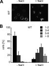
In vivo test for vacuole fragmentation. (A) Vacuole fragmentation in response to hypertonic stress. Wild-type cells (BJ3505) were grown in YPD at 25°C to logarithmic phase. Cells were stained with FM4-64, incubated for 10 min in the presence or absence of 0.4 M NaCl, and analyzed by fluorescence microscopy as described in Figure 1. (B) Quantification of vacuole morphology. The number of vacuolar vesicles per cell was determined, and cells were accordingly grouped into the indicated categories. For each experiment, 100 cells per strain and condition were analyzed. Three independent experiments were averaged, and standard deviations were calculated.
Figure 4.
Figure 4.
Fragmentation defects of V-ATPase mutants. Salt-induced vacuole fragmentation in V0 and V1 mutants. Yeasts were logarithmically grown in YPD, pH 5.5, at 25°C, stained with FM4-64, and subjected to a 10-min salt shock. Vacuolar morphology was quantified as described in Figure 1. (A) Wild-type, vma1Δ, vma2Δ, vma5Δ, vma3Δ, vma6Δ (in BY4741). (B) Wild-type and stv1Δ (in BJ3505). (C) vma4-1ts and its isogenic wild-type SF838-5A were treated and analyzed as described above, but the cells had been transferred to 37°C, or they were kept at 25°C for 40 min before the salt treatment.
Figure 5.
Figure 5.
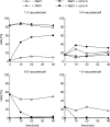
Proton translocation by the V-ATPase is indispensable for vacuole fragmentation. Effect of concanamycin A on in vivo vacuole fragmentation of wild-type cells (BJ3505). To block proton pumping by the V-ATPase, cells were incubated with 1 μM concanamycin A for 10, 20, or 40 min before hyperosmotic shock in 0.4 M NaCl.
Figure 6.
Figure 6.
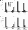
Suppression of fragmentation by concanamycin A results from reduced fission and not from enhanced fusion. Effect of concanamycin A on in vivo vacuole fragmentation of wild-type (A) and fusion-defective nyv1Δ cells (B; both in BJ3505). To block proton pumping by the V-ATPase, cells were incubated with 1 μM concanamycin A for 2 h before hyperosmotic shock in 0.4 M NaCl.
Figure 7.
Figure 7.
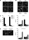
Epistasis of fragmentation and fusion defects. (A) Vacuole morphology of subunit a deletion mutants. Vacuolar membranes of wild-type, vph1Δ, stv1Δ, and stv1Δvph1Δ cells (all in BJ3505 background) were labeled with FM4-64 and visualized by fluorescence microscopy. (B) Effect of concanamycin A on vacuolar morphology of wild-type and vph1Δ cells. Cells were stained with FM4-64, and subsequently they were incubated for 2 h with 1 μM concanamycin A or control buffer (DMSO). Microscopy was as described in A. (C) Quantification of the experiment in B. (D) Vacuole morphology of nyv1Δvph1Δ cells after 2-h concanamycin A treatment as described in B.
Figure 8.
Figure 8.
Rescue of fusion-defective mutants by blocking proton pumping. (A) Vacuole structure in wild-type, vam3Δ, vma3Δ, and %vam3Δvma3Δ cells (all in BJ3505). Labeling and microscopy are as described in Figure 7A. (B–F) Effect of concanamycin A on the morphology of the indicated fusion-defective mutants (all in BJ3505 background). Labeling, concanamycin A treatment, and microscopy are as described in Figure 7B.
Figure 9.
Figure 9.
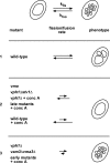
Schematic representation of the vacuolar fission-fusion equilibrium. Vacuolar morphology is determined by the equilibrium of the antagonistic processes of fission and fusion that constantly takes place with the specific rates kfis and kfus. Manipulations interfering with fission and/or fusion result in modified reaction rates. The relative changes in fission and fusion rates determine whether and to what extent the equilibrium point is shifted and hence the phenotypic outcome. Three cases can be distinguished: 1) Fission and fusion rates are balanced. Vacuole morphology is normal. 2) Fission is more strongly affected than fusion. As a result fusion prevails, leading to a cell with a single large vacuole. and 3) Fission is less strongly affected than fusion. As a consequence, fission outweighs fusion, resulting in a cell with fragmented vacuoles.

References

    1. Bayer M. J., Reese C., Buhler S., Peters C., Mayer A. Vacuole membrane fusion: V0 functions after trans-SNARE pairing and is coupled to the Ca2+-releasing channel. J. Cell Biol. 2003;162:211–222. - PMC - PubMed
    1. Bonangelino C. J., Nau J. J., Duex J. E., Brinkman M., Wurmser A. E., Gary J. D., Emr S. D., Weisman L. S. Osmotic stress-induced increase of phosphatidylinositol 3,5-bisphosphate requires Vac14p, an activator of the lipid kinase Fab1p. J. Cell Biol. 2002;156:1015–1028. - PMC - PubMed
    1. Brachmann C. B., Davies A., Cost G. J., Caputo E., Li J., Hieter P., Boeke J. D. Designer deletion strains derived from Saccharomyces cerevisiae S288C: a useful set of strains and plasmids for PCR-mediated gene disruption and other applications. Yeast. 1998;14:115–132. - PubMed
    1. Chan D. C. Mitochondria: dynamic organelles in disease, aging, and development. Cell. 2006;125:1241–1252. - PubMed
    1. Conradt B., Shaw J., Vida T., Emr S., Wickner W. In vitro reactions of vacuole inheritance in Saccharomyces cerevisiae. J. Cell Biol. 1992;119:1469–1479. - PMC - PubMed

Publication types

MeSH terms

LinkOut - more resources