Review
. 2007 Aug 1;4(4):418-30.
doi: 10.1513/pats.200703-041BR.

Endpoints for clinical trials in young children with cystic fibrosis

Affiliations
Review

Endpoints for clinical trials in young children with cystic fibrosis

Stephanie D Davis et al. Proc Am Thorac Soc. .

Abstract

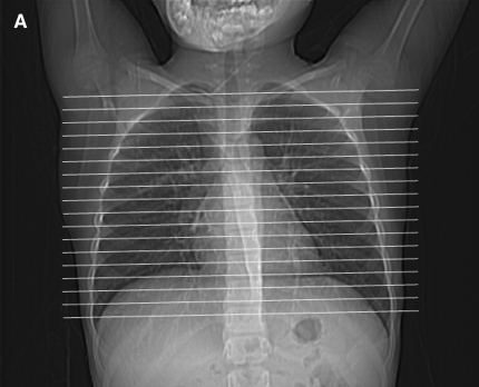
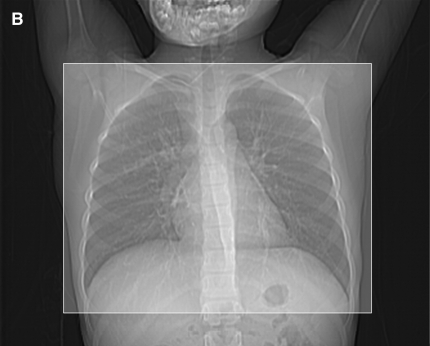
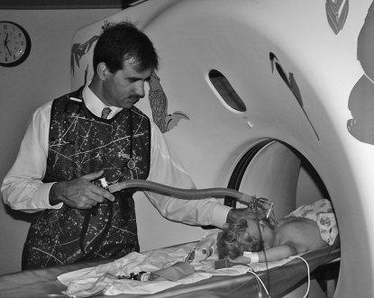
The availability of sensitive, reproducible, and feasible outcome measures for quantifying lung disease in children with cystic fibrosis (CF) younger than 6 years is critical to the conduct of clinical trials in this important population. Historically, identifying and quantifying the presence of lung disease in very young children with CF was hampered by a lack of reproducible measures of lung function or lung pathology. Over the past 10 years, significant progress has led to physiologic, anatomic, and bronchoscopic measures that may serve as endpoints for future intervention trials. These endpoints include infant and preschool lung function testing, computed tomography of the chest, and bronchoalveolar lavage markers of inflammation and infection. Much progress has occurred in standardizing lung function testing, which is essential for multicenter collaboration. Pulmonary exacerbation has the potential to serve as a clinical endpoint; however, there is currently no standardized definition in children with CF younger than 6 years. Further development of these outcomes measures will enable clinical trials in the youngest CF population with the objective of improving long-term prognosis.


Figures

<b>Figure 1.</b>
Figure 1.
(A) A sedated infant undergoing lung function testing with the raised volume rapid thoracoabdominal compression technique. (B) Schematic of the raised volume rapid thoracoabdominal compression technique. To initiate inflation, air is delivered from a compressed air source to the sedated infant through the inspiratory circuit. A pressure-relieve valve, located in the inspiratory circuit, is set at 30 cm H2O. During the inflation, the expiratory valve is closed, leading to inflation of the infant's lungs to 30 cm H2O. At the end of the inflation, the expiratory valve is then opened and the infant exhales passively. These inflation–passive exhalation maneuvers are repeated until a short respiratory pause is noted. Inflation is then repeated and the jacket is inflated (by opening a valve located between the jacket and the pressure reservoir) at end-inspiration to initiate the forced exhalation toward residual volume. The maneuver is repeated at increasing jacket pressures until flow limitation is achieved. (Reprinted by permission from Reference 48; and originally created by Marcus H. Jones, M.D., Ph.D.)
<b>Figure 2.</b>
Figure 2.
(A) Image shows mild bronchial wall thickening associated with bronchiectasis in the right upper lobe in a 2-year-old female with cystic fibrosis (CF). (B) Image displays multiple areas of air trapping (seen as dark areas), greatest in the right upper lobe in the same 2-year-old female with CF.
<b>Figure 3.</b>
Figure 3.
(A) High-resolution computed tomography (CT) scanning is a sampling technique that uses thin sections at intervals. Only the portion of the lungs indicated by the white lines is imaged. (B) Volumetric CT uses a helical technique to image a volume of tissue. The shaded square shows the area imaged using a volumetric technique.
<b>Figure 4.</b>
Figure 4.
Controlled ventilation computed tomography scanning. The respiratory circuit includes a pop-off valve that is set to 25 cm H2O pressure. The operator closes the circuit with his right thumb to administer positive pressure and augment the patient's inspiration. Several augmented inspirations cause a respiratory pause during which imaging is performed.

References

    1. Hammer J, Eber E, editors. Paediatric pulmonary function testing. Basel: Karger; 2005.
    1. Gappa M, Ranganathan S, Stocks J. Lung function testing in infants with cystic fibrosis: lessons from the past and future directions. Pediatr Pulmonol 2001;32:228–245. - PubMed
    1. Beardsmore CS, Bar-Yishay E, Maayan C, Yahav Y, Katznelson D, Godfrey S. Lung function in infants with cystic fibrosis. Thorax 1988;43:545–551. - PMC - PubMed
    1. Tepper RS, Montgomery GL, Ackerman V, Eigen H. Longitudinal evaluation of pulmonary function in infants and very young children with cystic fibrosis. Pediatr Pulmonol 1993;16:96–100. - PubMed
    1. Tepper RS, Hiatt P, Eigen H, Scott P, Grosfeld J, Cohen M. Infants with cystic fibrosis: pulmonary function at diagnosis. Pediatr Pulmonol 1988;5:15–18. - PubMed

Publication types