

A microarray analysis of retinal transcripts that are controlled by image contrast in mice
- PMID: 17653032
- PMCID: PMC2774458
A microarray analysis of retinal transcripts that are controlled by image contrast in mice
Abstract
Purpose: The development of myopia is controlled by still largely unknown retinal signals. The aim of this study was to investigate the changes in retinal mRNA expression after different periods of visual deprivation in mice, while controlling for retinal illuminance.
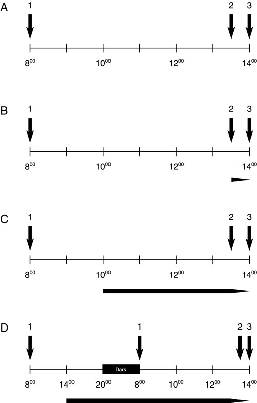
Methods: Each group consisted of three male C57BL/6 mice. Treatment periods were 30 min, 4 h, and 6+6 h. High spatial frequencies were filtered from the retinal image by frosted diffusers over one eye while the fellow eyes were covered by clear neutral density (ND) filters that exhibited similar light attenuating properties (0.1 log units) as the diffusers. For the final 30 min of the respective treatment period mice were individually placed in a clear Perspex cylinder that was positioned in the center of a rotating (60 degrees) large drum. The inside of the drum was covered with a 0.1 cyc/degree vertical square wave grating. This visual environment was chosen to standardize illuminances and contrasts seen by the mice. Labeled cRNA was prepared and hybridized to Affymetrix GeneChip Mouse Genome 430 2.0 arrays. Alterations in mRNA expression levels of candidate genes with potential biological relevance were confirmed by semi-quantitative real-time reverse transcription polymerase chain reaction (RT-PCR).
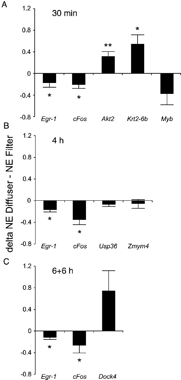
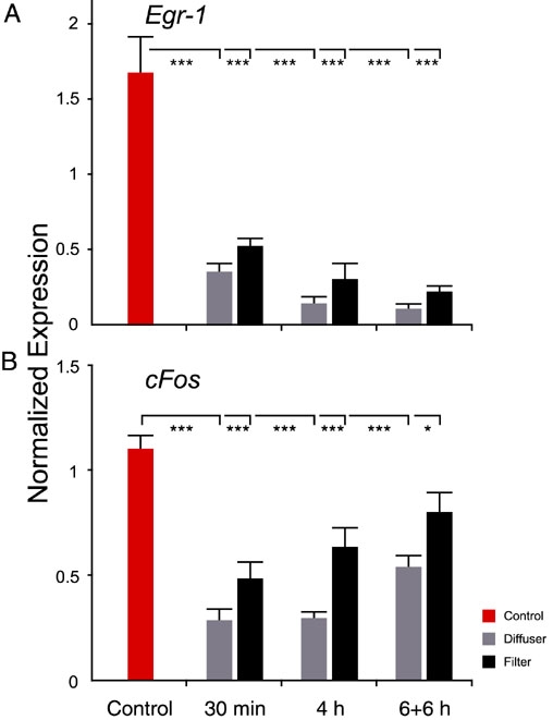
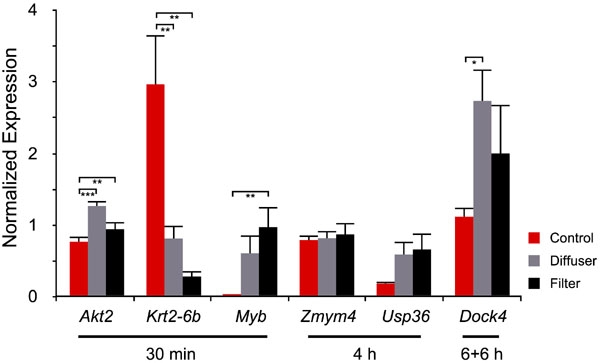
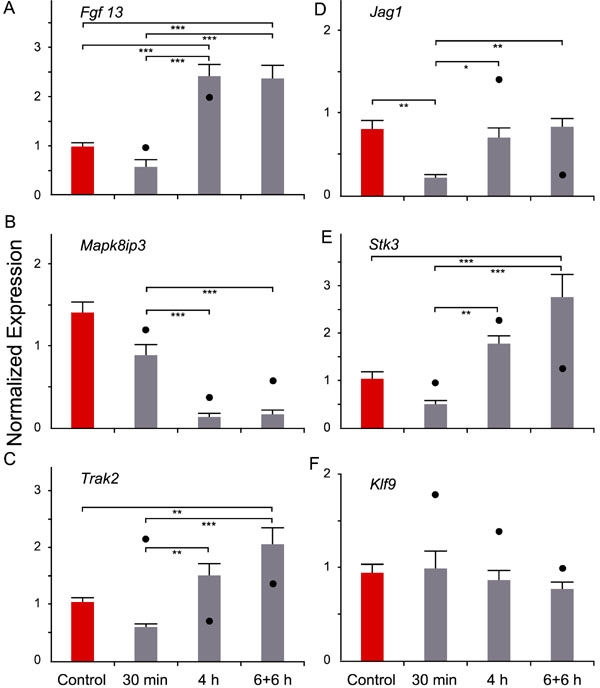
Results: In all groups, Egr-1 mRNA expression was reduced in diffuser-treated eyes. Furthermore, the degradation of the spatial frequency spectrum also changed the cFos mRNA level, with reduced expression after 4 h of diffuser treatment. Other interesting candidates were Akt2, which was up-regulated after 30 min of deprivation and Mapk8ip3, a neuron specific JNK binding and scaffolding protein that was temporally regulated in the diffuser-treated eyes only.
Conclusions: The microarray analysis demonstrated a pattern of differential transcriptional changes, even though differences in the retinal images were restricted to spatial features. The candidate genes may provide further insight into the biochemical short-term changes following retinal image degradation in mice. Because deprivation of spatial vision leads to increased eye growth and myopia in both animals and humans, it is believed some of the identified genes play a role in myopia development.
Figures
References
-
- Dirani M, Chamberlain M, Garoufalis P, Chen C, Guymer RH, Baird PN. Refractive errors in twin studies. Twin Res Hum Genet. 2006;9:566–72. - PubMed
-
- Wallman J, Turkel J, Trachtman J. Extreme myopia produced by modest change in early visual experience. Science. 1978;201:1249–51. - PubMed
-
- Hodos W, Kuenzel WJ. Retinal-image degradation produces ocular enlargement in chicks. Invest Ophthalmol Vis Sci. 1984;25:652–9. - PubMed
-
- Schaeffel F, Howland HC. Visual optics in normal and ametropic chickens. Clin Vis Sci. 1988;3:83–98.
-
- Sherman SM, Norton TT, Casagrande VA. Myopia in the lid-sutured tree shrew (Tupaia glis). Brain Res. 1977;124:154–7. - PubMed
Publication types
MeSH terms
Substances
LinkOut - more resources
Full Text Sources
Research Materials
Miscellaneous