

Clinicopathological and biological significance of mitotic centromere-associated kinesin overexpression in human gastric cancer
- PMID: 17653072
- PMCID: PMC2360338
- DOI: 10.1038/sj.bjc.6603905
Clinicopathological and biological significance of mitotic centromere-associated kinesin overexpression in human gastric cancer
Abstract
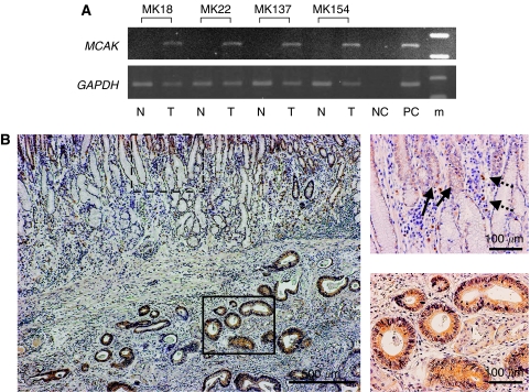
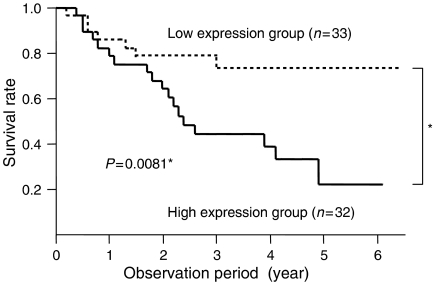
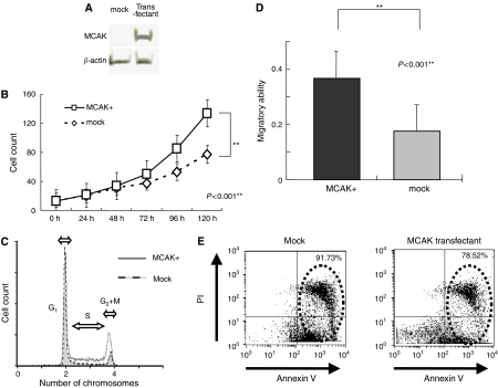
Mitotic centromere-associated kinesin (MCAK) is a microtubule (MT) depolymerase necessary for ensuring proper kinetochore MT attachment during spindle formation. To determine MCAK expression status and its clinicopathological significance, real-time reverse transcriptase-polymerase chain reaction was used in 65 cases of gastric cancer. MCAK gene expression in cancer tissue was significantly higher than expression in non-malignant tissue (P<0.05). Elevated MCAK expression was significantly associated with lymphatic invasion (P=0.01) and lymph node metastasis (P=0.04). Furthermore, patients with high MCAK expression had a significantly poorer survival rate than those with low MCAK expression (P=0.008). Immunohistochemical study revealed that expression of MCAK was primarily observed in cancer cells. Additionally, a gastric cancer cell line (AZ521) that stably expressed MCAK was established and used to investigate the biological effects of the MCAK gene. In vitro results showed that cells transfected with MCAK had a high rate of proliferation (P<0.001) and increased migratory ability (P<0.001) compared to mock-transfected cells. This study demonstrated that elevated expression of MCAK may be associated with lymphatic invasion, lymph node metastasis, and poor prognosis. These characteristics may be due in part to the increased proliferative and migratory ability of cells expressing MCAK.
Figures
Similar articles
-
Mitotic centromere-associated kinesin is a novel marker for prognosis and lymph node metastasis in colorectal cancer.Br J Cancer. 2008 Jun 3;98(11):1824-9. doi: 10.1038/sj.bjc.6604379. Epub 2008 May 27. Br J Cancer. 2008. PMID: 18506187 Free PMC article.
-
Relation between the expression of mitotic centromere-associated kinesin and the progression of squamous cell carcinoma of the tongue.Oral Surg Oral Med Oral Pathol Oral Radiol. 2014 Mar;117(3):353-60. doi: 10.1016/j.oooo.2013.11.488. Epub 2013 Nov 20. Oral Surg Oral Med Oral Pathol Oral Radiol. 2014. PMID: 24445227
-
Relationship between expression of IGFBP7 and clinicopathological variables in gastric cancer.J Clin Pathol. 2015 Oct;68(10):795-801. doi: 10.1136/jclinpath-2015-202987. Epub 2015 Jun 4. J Clin Pathol. 2015. PMID: 26043748
-
Elevated plasma osteopontin associated with gastric cancer development, invasion and survival.Gut. 2007 Jun;56(6):782-9. doi: 10.1136/gut.2006.109868. Epub 2006 Dec 5. Gut. 2007. PMID: 17148500 Free PMC article.
-
Molecular insight into the regulation and function of MCAK.Crit Rev Biochem Mol Biol. 2015 Jul-Aug;51(4):228-45. doi: 10.1080/10409238.2016.1178705. Epub 2016 May 5. Crit Rev Biochem Mol Biol. 2015. PMID: 27146484 Review.
Cited by
-
Genome-wide binding analysis unveils critical implication of B-Myb-mediated transactivation in cancers.Int J Biol Sci. 2024 Sep 3;20(12):4691-4712. doi: 10.7150/ijbs.92607. eCollection 2024. Int J Biol Sci. 2024. PMID: 39309447 Free PMC article.
-
Evaluation of public cancer datasets and signatures identifies TP53 mutant signatures with robust prognostic and predictive value.BMC Cancer. 2015 Mar 26;15:179. doi: 10.1186/s12885-015-1102-7. BMC Cancer. 2015. PMID: 25886164 Free PMC article.
-
Centrosome clustering and chromosomal (in)stability: a matter of life and death.Mol Oncol. 2011 Aug;5(4):324-35. doi: 10.1016/j.molonc.2011.05.003. Epub 2011 May 19. Mol Oncol. 2011. PMID: 21646054 Free PMC article. Review.
-
Mitotic centromere-associated kinesin is a novel marker for prognosis and lymph node metastasis in colorectal cancer.Br J Cancer. 2008 Jun 3;98(11):1824-9. doi: 10.1038/sj.bjc.6604379. Epub 2008 May 27. Br J Cancer. 2008. PMID: 18506187 Free PMC article.
-
Predictive and Prognostic Relevance of Tumor-Infiltrating Immune Cells: Tailoring Personalized Treatments against Different Cancer Types.Cancers (Basel). 2024 Apr 23;16(9):1626. doi: 10.3390/cancers16091626. Cancers (Basel). 2024. PMID: 38730579 Free PMC article. Review.
References
-
- Adrain C, Slee EA, Harte MT, Martin SJ (1999) Regulation of apoptotic protease activating factor-1 oligomerization and apoptosis by the WD-40 repeat region. J Biol Chem 274: 20855–20860 - PubMed
-
- Albini A, Iwamoto Y, Kleinman HK, Martin GR, Aaronson SA, Kozlowski JM, McEwan RN (1987) A rapid in vitro assay for quantitating the invasive potential of tumor cells. Cancer Res 47: 3239–3245 - PubMed
-
- Aoki S, Ohta K, Yamazaki T, Sugawara F, Sakaguchi K (2005) Mammalian mitotic centromere-associated kinesin (MCAK): a new molecular target of sulfoquinovosylacylglycerols novel antitumor and immunosuppressive agents. FEBS J 272: 2132–2140 - PubMed
-
- Bieche I, Olivi M, Champeme MH, Vidaud D, Lidereau R, Vidaud M (1998b) Novel approach to quantitative polymerase chain reaction using real-time detection: application to the detection of gene amplification in breast cancer. Int J Cancer 78: 661–666 - PubMed
Publication types
MeSH terms
Substances
LinkOut - more resources
Full Text Sources
Other Literature Sources
Medical
Research Materials
