. 2007 Oct;15(10):1872-80.
doi: 10.1038/sj.mt.6300270. Epub 2007 Jul 24.

A study of the role of nell-1 gene modified goat bone marrow stromal cells in promoting new bone formation

Affiliations

A study of the role of nell-1 gene modified goat bone marrow stromal cells in promoting new bone formation

Tara Aghaloo et al. Mol Ther. 2007 Oct.

Abstract

Nell-1 is a recently discovered secreted protein with the capacity to promote osteoblastic calvarial cell differentiation and mineralization and induce calvarial bone overgrowth and regeneration in various rodent models. However, the extent of Nell-1 osteoinductivity in large animal cells remains unknown. The objective of the study was to evaluate the feasibility of adenoviral encoding Nell-1 (AdNell-1) gene transfer into primary adult goat bone marrow stromal cells (BMSCs) in vitro and in vivo and to compare the osteoinductive effects with those produced by bone morphogenetic protein-2 (BMP-2), a well established osteoinductive molecule currently utilized for regional gene therapy. AdNell-1-transduced BMSCs expressed Nell-1 protein and underwent osteoblastic differentiation within 2 weeks in vitro, which is comparable to AdBMP-2. After intramuscular injection of nude mice, the AdNell-1- and AdBMP-2-transduced BMSCs revealed new bone formation, while untransduced or AdLacZ-transduced BMSCs showed mainly fibrotic tissue proliferation. At 4 weeks, BMP-2 induced significantly larger bone mass with a mature bone margin and central cavity filled with primarily fatty marrow tissue. Nell-1 samples had significantly less bone mass but were histologically similar to newly formed trabecular bone mixed with chondroid bone-like areas verified by type X collagen (ColX) immunohistochemistry. This distinct difference in histomorphology from the bone mass induced by BMP-2 suggests that there is a potential clinical role/advantage for Nell-1 in skeletal tissue engineering and regeneration.


Figures

Figure 1
Figure 1. Gene transduction and the effects on bone marrow stromal cell (BMSC) proliferation
(a) A multiplicity of infection of 50 pfu/cell achieved high transfer efficiency above 70% 3 days after AdLacZ transduction of goat BMSCs. Positive areas with X-gal staining are in blue. (b-d) Cellular morphology after transduction with AdNell-1, AdBMP-2, or untransduced control cells. Original magnification ×200. (e) Western blot probed with antibodies against Nell-1 and β-actin for confirmation of Nell-1 protein expression 3 days after gene transduction. (f) Effects of AdNell-1, AdBMP-2, and AdLacZ infection on BMSC proliferation. A growth curve represents cellular proliferation after transduction with AdNell-1, AdBMP-2, or AdLacZ. Data points represent the mean ± SD. A significant decrease in cell number after transduction with AdNell-1 compared to AdLacZ at days 3, 4, and 5; *P < 0.05. No significant difference was detected between AdBMP-2 and AdLacZ groups; P > 0.05.
Figure 2
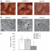
Figure 2. In vitro analysis of osteoblastic differentiation after bone marrow stromal cell transduction with AdNell-1, AdBMP-2, and AdLacZ
(a) alkaline phosphatase expression 12 days after gene transduction. (b) von Kossa assay comparing calcium nodules between AdNell-1-, AdBMP-2-, and AdLacZ-transduced cells 14 days after gene transfer. (c) Quantitative analysis of calcium nodules present in the three groups. A significant increase is seen in the AdNell-1 and AdBMP-2 groups when compared with the AdLacZ group; *P < 0.05, but no significant difference is seen between the AdNell-1 and AdBMP-2 groups; P > 0.05.
Figure 3
Figure 3. Histologic analysis of 2-week tissues in vivo
(a) AdNell-1-transduced bone marrow stromal cell (BMSC) injection sites showing cartilage (arrows) and osseous tissue (arrowheads) with the presence of chondroid matrix (CM) and bone marrow; hematoxylin and eosin (H&E) staining. (b) AdBMP-2 transduced BMSC injection sites showing osseous tissue intramuscularly (arrowheads) with cartilaginous tissue (arrows); H&E staining. (c) AdLacZ-transduced BMSC injection sites showing a small locus of cartilaginous (arrows) and fibroblastic (arrowheads) tissue; H&E staining. (d-f) Alcian blue staining on corresponding tissue sections of a-c to confirm the presence of cartilaginous tissue (blue) Original magnification for all figures: ×100.
Figure 4
Figure 4. Radiographic, microCT, and histologic evidence of bone formation after 4 weeks in vivo
(a) Plain radiography shows a large, defined radio-opaque mass (white arrow) with density similar to iliac bone, representing bone formation in the AdNell-1-transduced bone marrow stromal cell (BMSC) injection site on the left side as compared to the AdLacZ-treated right side. (b) An even larger radio-opaque mass with less dense bone compared to iliac bone is seen on the AdBMP-2-transduced BMSC injection site (white arrow). (c) 3D microCT analysis better demonstrates the bone mass seen in the AdNell-1 treated site (white arrow) similar to native bone, that could not be detected in AdLacZ sites. (d) 3D microCT image of a large bony nodule in the AdBMP-2-treated site (white arrow). (e) When the microCT image is bisected, a radio-dense mass is seen in the AdNell-1 treated site, (f) compared to a hollow cavity with only an outer bony shell with interspersed small bone trabeculae in the AdBMP-2-treated site. (g) Hematoxylin and eosin (H&E) histology shows the typical bony morphology in the AdNell-1 specimen compared to (h) a fatty marrow cavity with an outer bony surface and few bone trabeculae in the AdBMP-2 specimen (arrows). (i, l, o) H&E histological analysis (i) and immunostaining for Nell-1 (l) and bone morphogenetic protein-2 (BMP-2) (o) of in vivo bone formation in AdNell-1-transduced BMSC injection sites. Mature bone formation (arrows) with Haversian systems and marrow cavities with some fat cells are seen. Areas of chondroid bone matrix are also seen (CM). Nell-1 immunohistochemistry demonstrating positive brown staining in osteocytes (arrows) and some bone marrow cells (arrowheads). BMP-2 immunohistochemistry demonstrates low level staining only in new bone forming areas (arrows) and bone marrow cells (arrowheads). (j, m, p) H&E histology (j) and immunostaining for Nell-1 (m) and BMP-2 (p) of AdBMP-2-transduced BMSC injection sites demonstrates more mature but thinner trabecular bone scattered within a large amount of bone marrow in a big cavity filled with mostly fatty tissue, without obvious evidence of cartilage or chondroid matrix. Nell-1 immunohistochemistry shows no obvious staining, while BMP-2 immunohistochemistry demonstrates a strong BMP-2 expression in both the bone (arrows) and surrounding fibroblastic tissue (arrowheads). (k) Most of the AdLacZ-transduced BMSC injection sites show only fibroblastic tissue (arrows); (n) Frozen sections stained with X-gal after 4 weeks in AdLacZ-transduced BMSC injection sites (arrows). Original magnification for figure g and h: ×40; i, j, and k: ×100; l, m, n, o, and p: ×200).
Figure 5
Figure 5. Immunohistochemical analysis of intramuscular bone and cartilage formed in AdNell-1-, AdBMP-2-, or AdLacZ-transduced bone marrow stromal cell (BMSC) injection sites after 4 weeks in vivo
(a) The AdNell-1 group demonstrates significant bone matrix formation (BM) and bone marrow cavities (M) with some areas of immature bone containing large cells resembling chondrocytes (black arrows). (b) The AdBMP-2 group shows more mature bone matrix formation (BM) with marrow cavities (M) mainly filled with fatty tissue. (c) The AdLacZ group shows primarily cartilage (C) with some bone formation at the periphery (arrows and arrow heads) and marrow cavities (M). (d, e, f) Alcian blue better demonstrates the cartilage present in the corresponding tissue sections of figure a, b, and c. (g) Sox9 immunohistochemistry shows minimal staining in the AdNell-1 sites (arrows), (h) light staining in the AdBMP-2 sites, (i) but abundant positive brown staining localized in the nuclei in the AdLacZ sites (arrows). (j) Type X collagen (ColX) immunohistochemistry shows positive staining throughout the extracellular matrix and the hypertrophic chondrocytes (arrows) in the AdNell-1 sites. (k) In the AdBMP-2 sites, positive staining appears only in the extracellular matrix. (l) Isolated areas of positive staining for ColX were observed in the periphery of the cartilage where bone formation occurred in AdLacZ sites (arrows). Original magnification for all figures: ×200.

References

    1. Bauer TW, Muschler GF. Bone graft materials. An overview of the basic science. Clin Orthop Relat Res. 2000;371:10–27. - PubMed
    1. King GN. The importance of drug delivery to optimize the effects of bone morphogenetic proteins during periodontal regeneration. Curr Pharm Biotechnol. 2001;2:131–142. - PubMed
    1. Yoneda M, Terai H, Imai Y, Okada T, Nozaki K, Inoue H, et al. Repair of an intercalated long bone defect with a synthetic biodegradable bone-inducing implant. Biomaterials. 2005;26:5145–5152. - PubMed
    1. Govender S, Csimma C, Genant HK, Valentin-Opran A, Amit Y, Arbel R, et al. Recombinant human bone morphogenetic protein-2 for treatment of open tibial fractures: a prospective, controlled, randomized study of four hundred and fifty patients. J Bone Joint Surg Am. 2002;84-A:2123–2134. - PubMed
    1. Johnson EE, Urist MR. Human bone morphogenetic protein allografting for reconstruction of femoral nonunion. Clin Orthop Relat Res. 2000;371:61–74. - PubMed

Publication types

Substances

LinkOut - more resources