

Analysis and simulation of progressive adolescent scoliosis by biomechanical growth modulation
- PMID: 17653775
- PMCID: PMC2078290
- DOI: 10.1007/s00586-007-0442-7
Analysis and simulation of progressive adolescent scoliosis by biomechanical growth modulation
Abstract
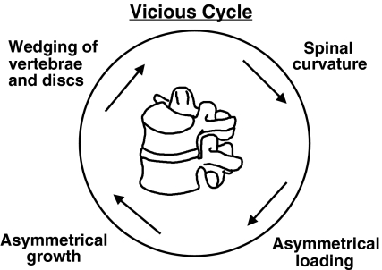
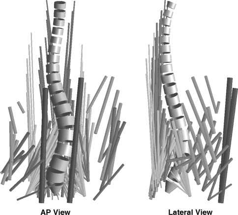
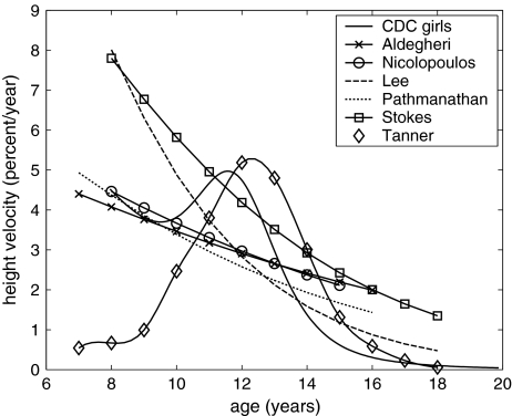
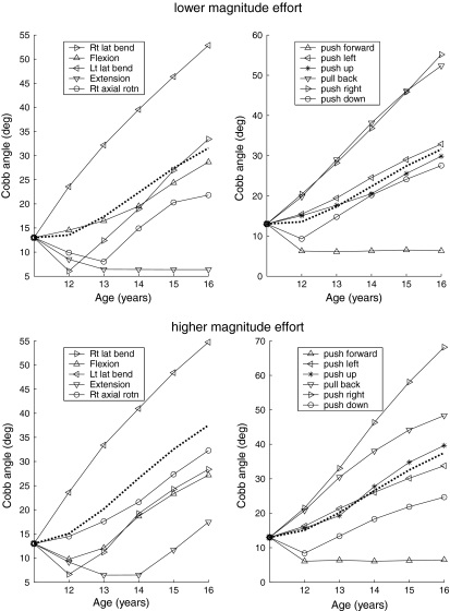
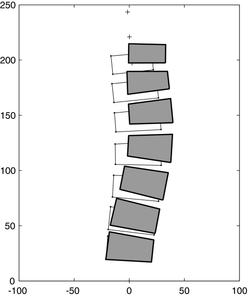
Scoliosis is thought to progress during growth because spinal deformity produces asymmetrical spinal loading, generating asymmetrical growth, etc. in a 'vicious cycle.' The aim of this study was to test quantitatively whether calculated loading asymmetry of a spine with scoliosis, together with measured bone growth sensitivity to altered compression, can explain the observed rate of scoliosis progression in the coronal plane during adolescent growth. The simulated spinal geometry represented a lumbar scoliosis of different initial magnitudes, averaged and scaled from measurements of 15 patients' radiographs. Level-specific stresses acting on the vertebrae were estimated for each of 11 external loading directions ('efforts') from published values of spinal loading asymmetry. These calculations assumed a physiologically plausible muscle activation strategy. The rate of vertebral growth was obtained from published reports of growth of the spine. The distribution of growth across vertebrae was modulated according to published values of growth sensitivity to stress. Mechanically modulated growth of a spine having an initial 13 degrees Cobb scoliosis at age 11 with the spine subjected to an unweighted combination of eleven loading conditions (different effort direction and magnitude) was predicted to progress during growth. The overall shape of the curve was retained. The averaged final lumbar spinal curve magnitude was 32 degrees Cobb at age 16 years for the lower magnitude of effort (that produced compressive stress averaging 0.48 MPa at the curve apex) and it was 38 degrees Cobb when the higher magnitudes of efforts (that produced compressive stress averaging 0.81 MPa at the apex). An initial curve of 26 degrees progressed to 46 degrees and 56 degrees, respectively. The calculated stresses on growth plates were within the range of those measured by intradiscal pressures in typical daily activities. These analyses predicted that a substantial component of scoliosis progression during growth is biomechanically mediated. The rationale for conservative management of scoliosis during skeletal growth assumes a biomechanical mode of deformity progression (Hueter-Volkmann principle). The present study provides a quantitative basis for this previously qualitative hypothesis. The findings suggest that an important difference between progressive and non-progressive scoliosis might lie in the differing muscle activation strategies adopted by individuals, leading to the possibility of improved prognosis and conservative or less invasive interventions.
Figures
References
-
- Aldegheri R, Agostini S. A chart of anthropometric values. J Bone Joint Surg Br. 1993;75(1):86–88. - PubMed
-
- Andersson GBJ, Chaffin DB, Pope MH. Occupational biomechanics of the lumbar spine. In: Pope MH, Frymoyer JW, Andersson G, editors. Occupational low back pain. New York: Praeger Scientific; 1984. p. 45.
-
- Cassella MC, Hall JE. Current treatment approaches in the nonoperative and operative management of adolescent idiopathic scoliosis. Phys Ther. 1991;71:897–909. - PubMed
-
- CDC Growth Charts: United States. STATAGE.XLS: Stature-for-age charts, 2 to 20 years. http://www.cdc.gov/nchs/about/major/nhanes/growthcharts/datafiles.htm. Accessed 12 Apr 2007
-
- Dickson RA, Deacon P. Annotation: spinal growth. J Bone Joint Surg Br. 1987;69:690–692. - PubMed
Publication types
MeSH terms
Grants and funding
LinkOut - more resources
Full Text Sources
Medical
Miscellaneous
