. 2008 Feb;214(2):389-95.
doi: 10.1002/jcp.21209.

Tissue stretch decreases soluble TGF-beta1 and type-1 procollagen in mouse subcutaneous connective tissue: evidence from ex vivo and in vivo models

Affiliations

Tissue stretch decreases soluble TGF-beta1 and type-1 procollagen in mouse subcutaneous connective tissue: evidence from ex vivo and in vivo models

Nicole A Bouffard et al. J Cell Physiol. 2008 Feb.

Abstract

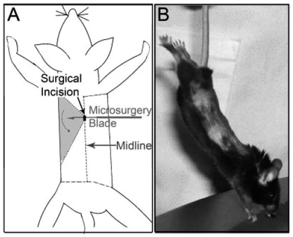
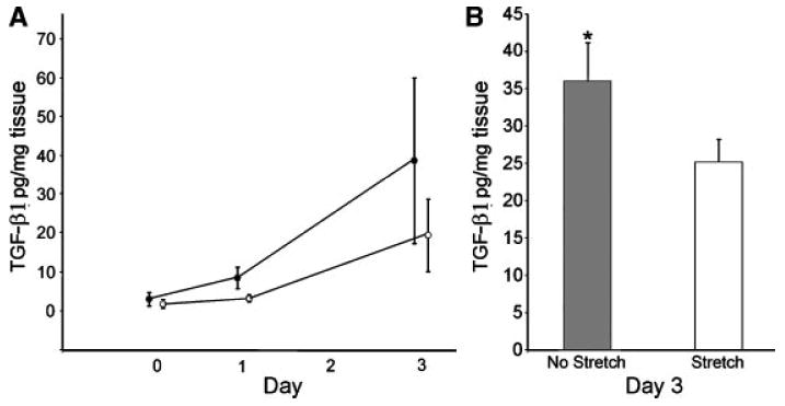
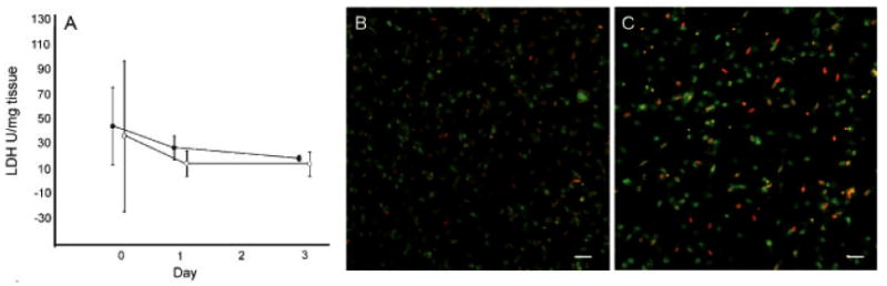
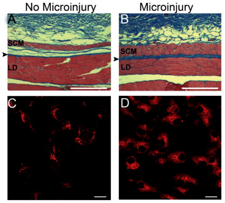
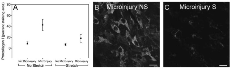
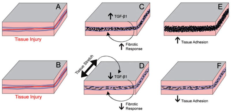
Transforming growth factor beta 1 (TGF-beta1) plays a key role in connective tissue remodeling, scarring, and fibrosis. The effects of mechanical forces on TGF-beta1 and collagen deposition are not well understood. We tested the hypothesis that brief (10 min) static tissue stretch attenuates TGF-beta1-mediated new collagen deposition in response to injury. We used two different models: (1) an ex vivo model in which excised mouse subcutaneous tissue (N = 44 animals) was kept in organ culture for 4 days and either stretched (20% strain for 10 min 1 day after excision) or not stretched; culture media was assayed by ELISA for TGF-beta1; (2) an in vivo model in which mice (N = 22 animals) underwent unilateral subcutaneous microsurgical injury on the back, then were randomized to stretch (20-30% strain for 10 min twice a day for 7 days) or no stretch; subcutaneous tissues of the back were immunohistochemically stained for Type-1 procollagen. In the ex vivo model, TGF-beta1 protein was lower in stretched versus non-stretched tissue (repeated measures ANOVA, P < 0.01). In the in vivo model, microinjury resulted in a significant increase in Type-1 procollagen in the absence of stretch (P < 0.001), but not in the presence of stretch (P = 0.21). Thus, brief tissue stretch attenuated the increase in both soluble TGF-beta1 (ex vivo) and Type-1 procollagen (in vivo) following tissue injury. These results have potential relevance to the mechanisms of treatments applying brief mechanical stretch to tissues (e.g., physical therapy, respiratory therapy, mechanical ventilation, massage, yoga, acupuncture).


Figures

Fig. 1
Fig. 1
A: In vivo mouse microinjury procedure. A microsurgery blade is introduced subcutaneously via a 5 mm midline incision. The subcutaneous tissue layer is cut over a 1.5 cm × 1.5 cm area on one side of the back lateral to the midline with the other side serving as the control. B: Method used to induce tissue stretch in vivo. Mice are suspended by the tail such that their paws barely touch a surface slightly inclined relative to the vertical. The mice spontaneously extend their front and hind limbs, the distance between ipsilateral hip and shoulder joints becoming 20–30% greater than the resting distance.
Fig. 2
Fig. 2
Effect of tissue stretch on TGF-β1 protein ex vivo. A: Time course of TGF-β1 protein levels in the culture media for non-stretched (closed circle, N = 4) and stretched (open circle, N = 4) mouse subcutaneous tissue explants on days 0, 1, and 3 post-stretch (or no stretch). All tissue samples were excised and incubated for 24h prior to day 0.B:Levels of TGF-β1 protein in the culture media at day 3 for non-stretched and stretched sbcutaneous tissue samples (N = 36). Asterisk (*) indicates significant difference from stretched (P = 0.002). Error bars represent standard errors.
Fig. 3
Fig. 3
Ex vivo tissue injury and cell viability assessment. A: Time course of LDH concentration in the culture media (marker of cell death) for non-stretched (closed circle, n = 4) and stretched (open circle, n = 4) mouse subcutaneous tissue explants on days 0, 1, and 3 post-stretch (or no stretch). B,C: Confocal microscopy imaging of mouse subcutaneous tissue explants showing similar proportions of live (green) and dead (red) cells in non-stretched (A) versus stretched (B) tissue after 3 day incubation post-stretch (or no stretch). Images are projections of three-dimensional image stacks. Scale bars: 40 μm.
Fig. 4
Fig. 4
Mouse in vivo microinjury model. Effect of microinjury on mouse subcutaneous tissue newly formed collagen. A,B: Masson Trichrome (stains collagen blue) of paraffin-embedded histological sections cut perpendicular to the skin; arrowhead indicates subcutaneous tissue located between the subcutaneous muscle (SCM) and latissimus dorsi (LD) muscle. C,D: Immunohistochemical staining for Type-1 procollagen (marker of newly formed collagen) of dissected whole subcutaneous tissue mounts. Scale bars: 1 mm (A,B) and 40 μm (C,D).
Fig. 5
Fig. 5
Effect of tissue stretch in vivo on subcutaneous tissue Type-1 procollagen in mouse microinjury model. A: Mean ± SE procollagen percent staining area in non-injured versus injured sides, without stretch (N = 11) and with stretch (N = 10); B,C: Type-1 procollagen in non-stretched and stretched tissue (both injured). Scale bars, 40 μm.
Fig. 6
Fig. 6
Proposed model for healing of connective tissue injury in the absence (A,C,E) and presence (B,D,F) of tissue stretch. In this model, brief stretching of tissue beyond the habitual range of motion reduces soluble TGF-β1 levels (D) causing a decrease in the fibrotic response, less collagen deposition, and reduced tissue adhesion (F) compared with no stretch (E). Black lines represent newly formed collagen.

References

    1. Annes JP, Chen Y, Munger JS, Rifkin DB. Integrin alphaVbeta6-mediated activation of latent TGF-beta requires the latent TGF-beta binding protein-1. J Cell Biol. 2004;165:723–734. - PMC - PubMed
    1. Annes JP, Munger JS, Rifkin DB. Making sense of latent TGFbeta activation. J Cell Sci. 2003;116:217–224. - PubMed
    1. Arora PD, Narani N, McCulloch CA. The compliance of collagen gels regulates transforming growth factor-beta induction of alpha-smooth muscle actin in fibroblasts. Am J Pathol. 1999;154:871–882. - PMC - PubMed
    1. Atance J, Yost MJ, Carver W. Influence of the extracellular matrix on the regulation of cardiac fibroblast behavior by mechanical stretch. J Cell Physiol. 2004;200:377–386. - PubMed
    1. Balachandran K, Konduri S, Sucosky P, Jo H, Yoganathan AP. An ex vivo study of the biological properties of porcine aortic valves in response to circumferential cyclic stretch. Ann Biomed Eng. 2006;34:1655–1665. - PMC - PubMed

Publication types

MeSH terms

LinkOut - more resources