. 2007 Aug 21;46(33):9380-7.
doi: 10.1021/bi700578v. Epub 2007 Jul 27.

tRNase Z catalysis and conserved residues on the carboxy side of the His cluster

Affiliations

tRNase Z catalysis and conserved residues on the carboxy side of the His cluster

Shay Karkashon et al. Biochemistry. .

Abstract

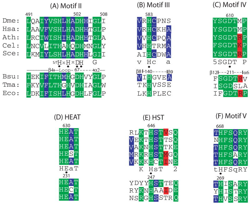
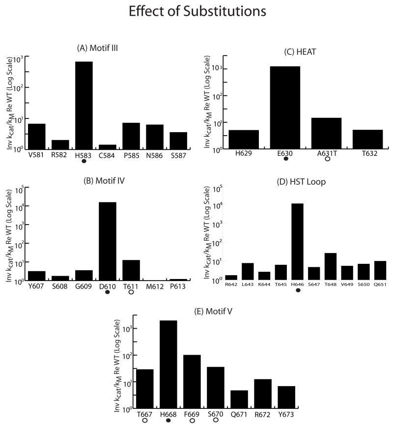
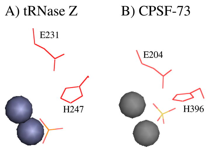
tRNAs are transcribed as precursors and processed in a series of required reactions leading to aminoacylation and translation. The 3'-end trailer can be removed by the pre-tRNA processing endonuclease tRNase Z, an ancient, conserved member of the beta-lactamase superfamily of metal-dependent hydrolases. The signature sequence of this family, the His domain (HxHxDH, Motif II), and histidines in Motifs III and V and aspartate in Motif IV contribute seven side chains for the coordination of two divalent metal ions. We previously investigated the effects on catalysis of substitutions in Motif II and in the PxKxRN loop and Motif I on the amino side of Motif II. Herein, we present the effects of substitutions on the carboxy side of Motif II within Motifs III, IV, the HEAT and HST loops, and Motif V. Substitution of the Motif IV aspartate reduces catalytic efficiency more than 10,000-fold. Histidines in Motif III, V, and the HST loop are also functionally important. Strikingly, replacement of Glu in the HEAT loop with Ala reduces efficiency by approximately 1000-fold. Proximity and orientation of this Glu side chain relative to His in the HST loop and the importance of both residues for catalysis suggest that they function as a duo in proton transfer at the final stage of reaction, characteristic of the tRNase Z class of RNA endonucleases.


Figures

Figure 1
Figure 1. Multiple sequence alignment of conserved loops involved in tRNase Z catalysis
A)Motif II; B) Motif III; C) Motif IV; D) HEAT Loop; E) HST Loop; F) Motif V. The top five aligned sequences are tRNase ZLs from Drosophila melanogaster, Homo sapiens, Arabidopsis thaliana, Caenorhabditis elegans and Saccharomyces cerevisiae (accession numbers Q8MKW7, NP_060597.3, AAM51378, O4476 and NP013005.1, respectively). Numbering of selected residues above the panels is for fruit fly tRNase ZL (as previously described; Zareen et al., 2005) based on the presumed translation initiation at an internal methionine (r24). Spacing between the homology blocks presented is also conserved in the five tRNase ZLs. Designations below the tRNase ZL panels indicate the consensus and coloring of residues indicates the extent of homology (green: identical; red: conserved in most cases; purple: similar). 3s correspond to the key resdieues in each block (discussed in text). The bottom three sequences are tRNase ZSs from B. subtilis, T. maritima and E. coli (accession numbers P54548, NP_228673 and ZP_00726790, respectively). Residue numbers and designations above the bottom panels, based on Bsu tRNase ZS (de la Sierra Gallay et al., 2005), indicate the positions of the secondary structure elements within the loops. The 36 residue gap between Motif III-IV in tRNase ZL relative to tRNase ZS is due to the flexible domain which is present at this location in tRNase ZS and only in the amino half of tRNase ZL.
Figure 2
Figure 2. The Bacillis subtilis tRNase Z structure model (de la Sierra Gallay et al., 2005; PDB#1Y44), including Motif II and loops on the carboxy side of Motif II, displayed with PyMol (Delano, 2002)
A) Best view of the entire tRNase ZS structure, a homodimer. The binding subunit (blue, upper left) is characterized by the flexible domain (FD). The active subunit (red, lower right) is dominated by the active site (marked with a fortuitously bound phosphate, orange, in the position suggested by de la Sierra-Gallay et al to be occupied by the scissile phosphodiester linkage in the pre-tRNA substrate). The entrance to the active site appears to be covered by a flap (the PxKxRN loop; Zareen et al., 2006). (B) – (F) Views of each of the loops on the carboxy side of Motif II that are demonstrably important for catalysis. Motif II, two zinc ions (grey spheres) and the phosphate (orange) that marks the active site are included to provide a frame of reference. These models are presented on the same scale but have been rotated to most clearly display the loops. Relevant side chains are displayed as lines and are labeled with the single letter code for the amino acids (H – His; D – Asp; E – Glu). B) Motif III: His 140 (His 583 in D. melanogaster; see alignments in Fig. 1) and the ends of the loop are marked. C) Motif IV: Asp 211 (Asp 610 in Dme) and the ends of the loop are marked. D) HEAT: Side chains of His 230 (His 629 in Dme) and Glu 231 (Glu 630 in Dme) are displayed. Note that Glu 231 is pointing toward His 247 and the active site and His 230 is pointing away from the active site. E) The HST loop: His 247 (His 646 in Dme) is displayed and end residues are marked to establish orientation. F) Motif V: His 269 (His 668 in Dme) is displayed and ends are marked. G) A composite of the loops from (B) – (F). Motif II is at upper right. Zinc ions and phosphate are omitted. H) Catalytically important residues in tRNase Z. Motif II residues (His 63, His 65, Asp 67, His 68) are at lower left. Side chain of Glu 231 (Glu 630 in Dme) is pointing down from upper right, toward His 247. In all the detail views (B – G), a groove or channel occupied by two zincs and a phosphate appears to run between catalytically important signature residues of Motif II (HxHxDH) and the functional elements that approach from the other side (de la Sierra-Gallay et al., 2005).
Figure 3
Figure 3. Kinetics of tRNase Z processing
A) Canonical cloverleaf structure and 17 nt 3’ end trailer of nuclear-encoded fruit fly pre-tRNAArg(UCG) substrate (Acc. # AE003494, L09202, nt#1197–1269). The tRNase Z cleavage site following the discriminator base (G73) is marked with an arrow. B) Soluble baculovirus-expressed tRNase Z electrophoresed on a 10% polyacrylamide SDS gel. Wild type and the Motif III variants R582A, H583A and C584A were loaded in lanes 1–4, respectively, as designated below the lanes. M: Invitrogen Benchmark ladder was loaded in the marker lane. Marker sizes are indicated at left. tRNase Z, as designated at right, has an apparent molecular weight of ~90 kD. Protein loads were 2.5 μl of the first dilution (meant to be 200 ng/μL) in a series used in processing experiments (see below) and tRNase Z band intensities (relative to a known tRNase Z standard, not shown) were used to adjust the value used for enzyme concentration to calculate kcat from Vmax. C) Michaelis-Menten processing experiments. Reactions with wild type and variant tRNase Z at enzyme concentrations indicated above the panels (as determined from the protein gel lanes in B) were sampled after 5, 10 and 15 min of incubation and electrophoresed on 6% denaturing polyacrylamide gels (brackets below the gels). Reactions were performed with constant labeled substrate and with varying unlabelled substrate concentration over the range from 2–50 nM as indicated below the gel panels. Images were obtained from dried gels using a Typhoon scanner (GE Healthcare) and analyzed with Imagequant software. M – a labeled DNA marker with sizes as indicated at left. –Pre and –tRNA designations at right refer to the precursor and tRNA product (as in A above). D) Superimposed Eadie – Hofstee plots of V/[S] vs. V (values obtained from traces of gel lanes as in (C); see methods). Slope (−kM) for H583A is slightly lower than the others. Vmax (intercept in equations at right of plot) and [Enzyme] from panel (B) is used to obtain kcat (see Table 1).
Figure 4
Figure 4. Bar graphs illustrating the effects of substitutions in conserved regions on the C-side of Motif II
The inverse of kcat/kM relative to wild type (taken from column 5 in Table 1) is presented on a log scale. Conserved residues (taken from Figure 1A) which affect kcat/kM are designated with symbols (3 for intermediate reductions and 3 for the greatest reductions). A) The Motif III region; B) the Motif IV region; C) HEAT; D) the HST loop; E) the Motif V region.
Figure 5
Figure 5. A catalytically important conserved Glu – His contact
A) Glu 231 and His 247 are shown along with spheres corresponding to two zinc ions and a phosphate that marks the active site of tRNase Z (de la Sierra-Gallay et al., 2005; PDB#1Y44). B) The structurally similar placement of Glu 204 and His 396 in human CPSF 73, shown along with spheres corresponding to two zinc ions and a sulfate that marks the active site (Mandel et al., 2006; PDB#2I7T).

Similar articles

Cited by

References

    1. Callebaut I, Moshous D, Mornon J-P, de Villartay J-P. Metallo-3-lactamase fold within nucleic acids processing enzymes: the 3-CASP family. Nucleic Acids Res. 2002;30:3592–3601. - PMC - PubMed
    1. de la Sierra-Gallay IL, Pellegrini O, Condon C. Structural basis for substrate binding, cleavage and allostery in the tRNA maturase RNase Z. Nature. 2005;433:657–661. - PubMed
    1. de la Sierra-Gallay IL, Mathy N, Pellegrini O, Condon C. Structure of the ubiquitous 3′ processing enzyme RNase Z bound to transfer RNA. Nat Struct Mol Biol. 2006;13:376–377. - PubMed
    1. DeLano WL. The PyMOL Molecular Graphics System on World Wide Web. 2002. http://www.pymol.org.
    1. Dominski Z, Yang XC, Marzluff WF. The polyadenylation factor CPSF-73 is involved in histone-pre-mRNA processing. Cell. 2005;123:37–48. - PubMed

Publication types

LinkOut - more resources