Review
. 2007 Jul 26:2:34.
doi: 10.1186/1750-1172-2-34.

Sweet's syndrome--a comprehensive review of an acute febrile neutrophilic dermatosis

Affiliations
Review

Sweet's syndrome--a comprehensive review of an acute febrile neutrophilic dermatosis

Philip R Cohen. Orphanet J Rare Dis. .

Abstract

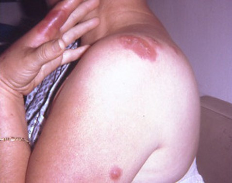
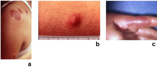
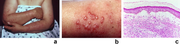
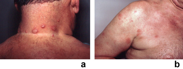
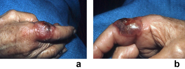
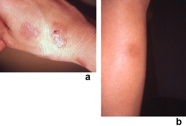
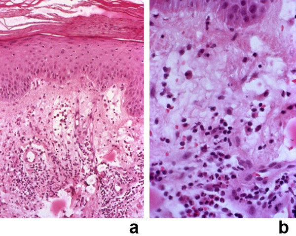
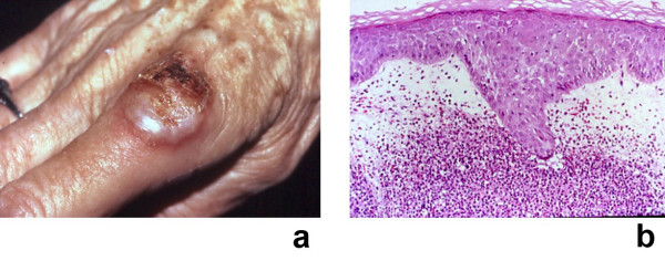
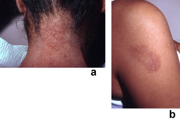
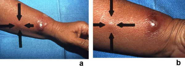
Sweet's syndrome (the eponym for acute febrile neutrophilic dermatosis) is characterized by a constellation of clinical symptoms, physical features, and pathologic findings which include fever, neutrophilia, tender erythematous skin lesions (papules, nodules, and plaques), and a diffuse infiltrate consisting predominantly of mature neutrophils that are typically located in the upper dermis. Several hundreds cases of Sweet's syndrome have been published. Sweet's syndrome presents in three clinical settings: classical (or idiopathic), malignancy-associated, and drug-induced. Classical Sweet's syndrome (CSS) usually presents in women between the age of 30 to 50 years, it is often preceded by an upper respiratory tract infection and may be associated with inflammatory bowel disease and pregnancy. Approximately one-third of patients with CSS experience recurrence of the dermatosis. The malignancy-associated Sweet's syndrome (MASS) can occur as a paraneoplastic syndrome in patients with an established cancer or individuals whose Sweet's syndrome-related hematologic dyscrasia or solid tumor was previously undiscovered; MASS is most commonly related to acute myelogenous leukemia. The dermatosis can precede, follow, or appear concurrent with the diagnosis of the patient's cancer. Hence, MASS can be the cutaneous harbinger of either an undiagnosed visceral malignancy in a previously cancer-free individual or an unsuspected cancer recurrence in an oncology patient. Drug-induced Sweet's syndrome (DISS) most commonly occurs in patients who have been treated with granulocyte-colony stimulating factor, however, other medications may also be associated with DISS. The pathogenesis of Sweet's syndrome may be multifactorial and still remains to be definitively established. Clinical and laboratory evidence suggests that cytokines have an etiologic role. Systemic corticosteroids are the therapeutic gold standard for Sweet's syndrome. After initiation of treatment with systemic corticosteroids, there is a prompt response consisting of dramatic improvement of both the dermatosis-related symptoms and skin lesions. Topical application of high potency corticosteroids or intralesional corticosteroids may be efficacious for treating localized lesions. Other first-line oral systemic agents are potassium iodide and colchicine. Second-line oral systemic agents include indomethacin, clofazimine, cyclosporine, and dapsone. The symptoms and lesions of Sweet's syndrome may resolved spontaneously, without any therapeutic intervention; however, recurrence may follow either spontaneous remission or therapy-induced clinical resolution.


Figures

Figure 1
Figure 1
Sweet's syndrome skin lesions in a woman with classical Sweet's syndrome. Cutaneous lesions of classical Sweet's syndrome on the left hand, left proximal arm and left shoulder in a 48-year-old woman with pyrexia, neutropenia, and a recent respiratory tract infection. (From [10] Cohen PR, Almeida L, Kurzrock R: Acute febrile neutrophilic dermatosis. Am Fam Physician 1989;39(3):199–204. Copyright 1989, Reprinted with permission from Academy of American Family Physicians, Leewood, Kansas.)
Figure 2
Figure 2
(a, b, and c). Tender, red Sweet's syndrome lesions in a woman with the classical form of the dermatosis. A closer view of the Sweet's syndrome lesions from the woman in Figure 1. The skin lesions improved rapidly after corticosteroid therapy was initiated. There is an erythematous plaque, 5 cm in diameter, with a pseudovesicular appearance on the left shoulder of the patient (a). A nodular lesion, 1 cm in diameter, is present on the lateral left arm (b). Painful, erythematous, pseudovesicular plaques of acute febrile neutrophilic dermatosis are present on the left hand (c). (From [10] Cohen PR, Almeida L, Kurzrock R: Acute febrile neutrophilic dermatosis. Am Fam Physician 1989;39(3):199–204. Copyright 1989, Reprinted with permission from Academy of American Family Physicians, Leewood, Kansas.)
Figure 3
Figure 3
(a, b, and c). Drug-induced Sweet's syndrome with photodistributed skin lesions. Photodistributed drug-induced Sweet's syndrome in a 50-year-old woman with trimethoprim-sulfamethoxazole-associated acute febrile neutrophilic dermatosis. Distant (a) and closer (b) views of Sweet's syndrome lesions located on the sun-exposed areas of the arms, hands, and upper chest are shown. The biopsy specimen (c) shows a confluent neutrophilic infiltrate in the reticular dermis and edema in the papillary dermis (hematoxylin and eosin, × 50). (From [13] Walker DC, Cohen PR: Trimethoprim-sulfamethoxazole-associated acute febrile neutrophilic dermatosis: case report and review of drug-induced Sweet's syndrome. J Am Acad Dermatol 1996;34:918–923. Copyright 1996, Reprinted with permission from the American Academy of Dermatology, Inc, Elsevier Ltd, Oxford, United Kingdom.)
Figure 4
Figure 4
(a and b). Recurrent Sweet's syndrome in a man with antiphospholipid syndrome. A 65-year-old white man with a 7-year history of recurrent Sweet's syndrome and antiphospholipid syndrome (initially manifested by multiple pulmonary emboli and subsequently managed with chronic coumadin therapy). Painful, red, biopsy-confirmed, pseudovesicular nodules and plaques of Sweet's syndrome are present on the posterior neck (a), chest, and upper arm (b). Although the Sweet's syndrome skin lesions resolved after the initiation of oral prednisone (60 mg/day), they recurred when the dose of the drug was subsequently tapered; potassium iodide, colchicine, dapsone, antimalarials, azathioprine, methotrexate, retinoids, and nonsteroidal anti-inflammatory agents were not effective as corticosteroid-sparing agents. (From [1] Cohen PR, Kurzrock R: Sweet's syndrome revisited: a review of disease concepts. Int J Dermatol 2003;42:761–778. Copyright 2003, Reprinted with permission from the International Society of Dermatology, Blackwell Publishing, Oxford, United Kingdom.)
Figure 5
Figure 5
(a and b). Radiotherapy-related Sweet's syndrome skin lesions in a woman with malignancy-associated Sweet's syndrome. Radiotherapy associated-exacerbated Sweet's syndrome in a patient with chronic lymphocytic leukemia and cutaneous squamous cell carcinoma. This biopsy-confirmed culture-negative, erythematous-based hemorrhagic, and vesicular-appearing Sweet's syndrome lesion extends from the dorsal (a) to the palmar (b) surface of the radial side of the right index finger and involves the skin between the metacarpal-phalangeal joint and the proximal interphalangeal joint of a 77-year-old woman. She has a prior medical history of hypothyroidism for which she receives daily Synthroid. Twenty-four months earlier, she had been diagnosed with chronic lymphocytic leukemia, which is adequately being managed with 2 mg of Myleran each day; her current white blood cell count of 51,800 cells/mm3 consists of 79% neutrophils, 15% bands, and 2% lymphocytes. More recently (3 months previously), a biopsy confirmed (at both initial microscopic evaluation and subsequent consultation pathology review) cutaneous squamous cell carcinoma involving the left index finger. She was started on an oral antibiotic (ciprofloxaxin), and radiotherapy (in fractionated doses over a period of 3 weeks) to the left index finger tumor was performed; during this treatment, a clinically similar-appearing lesion whose "presentation was suspicious for squamous cell carcinoma" began to develop on her right index finger. Based only on the lesion's morphologic characteristics, a single treatment with radiotherapy was also given; promptly thereafter, the right index finger lesion rapidly increased in size. Within a week, the lesion on the right index finger (as shown in a and b) had grown to a 4- × 4-cm pseudovesicular nodule that nearly involved the entire circumference of the digit and she was referred to the dermatology clinic. The lesion was painful and her leukocyte count was markedly elevated; however, she was (and had been) afebrile. Lesional biopsies for microscopic and culture evaluation were performed. Because the diagnosis of Sweet's syndrome was suspected, daily oral corticosteroid therapy with 60 mg prednisone was started. Oral cephalexin (250 mg 4 times each day for 7 days) and topical 2% mupirocin ointment (3 times each day) were also given. After a week of therapy, the lesion was greatly improved: it was no longer tender and it had decreased in size. Subsequently, the lesion completely resolved. The dose of prednisone was tapered during the next 5 weeks and then discontinued. (From [23] Cohen PR, Kurzrock R: Sweet's syndrome: a neutrophilic dermatosis classically associated with acute onset and fever. Clin Dermatol 2000;18:265–282. Copyright 2000, Reprinted with permission from Elsevier Ltd, Oxford, United Kingdom.)
Figure 6
Figure 6
(a and b). Clinical presentation of concurrent Sweet's syndrome and erythema nodosum. A 30-year-old woman had been receiving an oral contraceptive (0.3 mg norgestrel with 0.3 mg ethinyl estradiol) and an appetite suppressant (105 mg phendimetrazine tartrate) during the 5 months before these dermatoses appeared. An episode of recurrent herpes labialis involving her right upper lip also began 2 days prior to the onset of her skin lesions. Tender, erythematous plaques of biopsy-confirmed Sweet's syndrome are shown on the dorsal right hand and wrist; the suture indicates the biopsy site (a). A tender, erythematous nodule of biopsy confirmed erythema nodosum – which morphologically mimiced subcutaneous Sweet's syndrome – is shown on her right pretibial region (b). (From [21] Cohen PR, Holder WR, Rapini RP: Concurrent Sweet's syndrome and erythema nodosum: a report, world literature review and mechanism of pathogenesis. J Rheumatol 1992;5:814–820. Copyright 1992, Reprinted with permission from the Journal of Rheumatology, Toronto, Ontario.)
Figure 7
Figure 7
(a and b). Histopathology of a Sweet's syndrome lesion from a patient with concurrent erythema nodosum. Skin biopsy from one of the Sweet's syndrome plaques on the right dorsal wrist of a woman with concurrent biopsy-confirmed Sweet's sydrome and biopsy-confirmed erythema nodosum. The characteristic histopathologic features of Sweet's syndrome are observed at low (a) and higher (b) magnification: papillary dermal edema, swollen endothelial cells, and a diffuse infiltrate of predominantly neutrophils with leukocytoclasia. There is no evidence of vasculitis (hematoxylin and eosin, × 25 (a), × 100 (b)). (From [21] Cohen PR, Holder WR, Rapini RP: Concurrent Sweet's syndrome and erythema nodosum: a report, world literature review and mechanism of pathogenesis. J Rheumatol 1992;5:814–820. Copyright 1992, Reprinted with permission from the Journal of Rheumatology, Toronto, Ontario.)
Figure 8
Figure 8
(a and b). Malignancy-associated Sweet's syndrome in a woman with dermatosis-related laryngeal carcinoma. Solid tumor-associated Sweet's syndrome in a 69-year-old woman in whom a workup after the appearance of Sweet's syndrome lesions revealed an unsuspected recurrent squamous cell carcinoma of the larynx. A tender, erythematous-based, vesicular-appearing nodule of Sweet's syndrome with central ulceration and crust located proximally on the dorsum of the right second digit is shown (a). The histologic features of the Sweet's syndrome lesion revealed a neutrophilic dermatosis; the papillary dermis was edematous and contained a dense infiltrate of neutrophils (b) (hematoxylin and eosin, × 50). (From [362] Cohen PR, Holder WR, Tucker SB, Kono S, Kurzrock R: Sweet's syndrome in patients with solid tumors. Cancer 1993;72:2723–2731. Copyright 1993, Reprinted with permission from John Wiley & Sons, Inc., Hoboken, New Jersey.)
Figure 9
Figure 9
(a and b). Recurrent, oral corticosteroid-responsive, Sweet's syndrome lesions. A 46-year-old Indian woman with oral corticosteroid-responsive, tender erythematous, biopsy-confirmed plaques of recurrent Sweet's syndrome on the posterior neck (a) and right arm (b). Her laboratory evaluation was significant for the elevation of the erythrocyte sedimentation rate, liver function tests [serum glutamic-oxaloacetic transaminase (SGOT), serum glutamic-pyruvic transaminase (SGPT), and lactate dehydrogenase (LDH)], cytomegalovirus immunoglobulin G (IgG) antibody (consistent with previous exposure), hepatitis A IgG antibody (consistent with previous infection), and Epstein-Barr virus capsid IgG antibody (consistent with recent or past infection). (From [1] Cohen PR, Kurzrock R: Sweet's syndrome revisited: a review of disease concepts. Int J Dermatol 2003;42:761–778. Copyright 2003, Reprinted with permission from the International Society of Dermatology, Blackwell Publishing, Oxford, United Kingdom.)
Figure 10
Figure 10
(a and b). Sweet's syndrome with lesions distributed in a sporotrichoid pattern. A 53-year-old Hispanic woman presented to the emergency room with a recent episode of a "sore throat," fever, and a 2-day history of a swollen, tender left wrist accompanied by erythema that was beginning to extend proximally; a bacterial cellulitis was clinically suspected and she was started on oral antibiotics: double-strength trimethoprim-sulfamethoxazole twice daily and 400 mg of ofloxacin twice daily. She was initially seen in the dermatology clinic 9 days later; she was still febrile and her original lesion had developed into a painful larger pseudovesicular nodule on the radial side of her left wrist. In addition, distal (a) and closer (b) views show a smaller red dermal nodule (between arrows) that appeared on her left arm proximal to the original lesion. The clinical differential diagnosis included infections whose lesions demonstrated a sporotrichoid pattern (sporotrichosis and atypical mycobacterial infection) and Sweet's syndrome. Biopsies for microscopic and culture evaluation were performed. In addition to her antibiotics, the patient was started on oral saturated solution of potassium iodide (3 drops 3 times each day and increased by 1 drop each day to a final dose of 10 drops 3 times each day). The hematoxylin and eosin-stained sections from her biopsy showed a neutrophilic dermatosis; the bacterial, mycobacterial, and fungal cultures were negative for organisms. Within a few days after initiating treatment with potassium iodide, her symptoms resolved and her skin lesions began to improve. (From [23] Cohen PR, Kurzrock R: Sweet's syndrome: a neutrophilic dermatosis classically associated with acute onset and fever. Clin Dermatol 2000;18:265–282. Copyright 2000, Reprinted with permission from Elsevier Ltd, Oxford, United Kingdom.)

References

    1. Cohen PR, Kurzrock R. Sweet's syndrome revisited: a review of disease concepts. Int J Dermatol. 2003;42:761–778. doi: 10.1046/j.1365-4362.2003.01891.x. - DOI - PubMed
    1. Cohen PR. Sweet's syndrome. Orphanet Encyclopedia. 2003. http://www.orpha.net/data/patho/GB/uk-Sweet.pdf (accessed 25 July 2007).
    1. Cohen PR. Skin lesions of Sweet syndrome and its dorsal hand variant contain vasculitis: an oxymoron or an epiphenomenon? Arch Dermatol. 2002;138:400–403. doi: 10.1001/archderm.138.3.400. - DOI - PubMed
    1. Cohen PR. Subcutaneous Sweet's syndrome: a variant of acute febrile neutrophilic dermatosis that is included in the histologic differential diagnosis of neutrophilic panniculitis. J Am Acad Dermatol. 2005;52:927–928. doi: 10.1016/j.jaad.2005.03.001. - DOI - PubMed
    1. Cohen PR. Sweet's syndrome and relapsing polychondritis:is their appearance in the same patient a coincidental occurrence or a bonified association of these conditions? Int J Dermatol. 2004;43:772–777. doi: 10.1111/j.1365-4632.2004.02197.x. - DOI - PubMed

LinkOut - more resources