

Inhibition of APN/CD13 leads to suppressed progressive potential in ovarian carcinoma cells
- PMID: 17655775
- PMCID: PMC2000898
- DOI: 10.1186/1471-2407-7-140
Inhibition of APN/CD13 leads to suppressed progressive potential in ovarian carcinoma cells
Abstract
Background: Aminopeptidase N (APN/CD13), a 150-kDa metalloprotease, is a multifunctional cell surface aminopeptidase with ubiquitous expression. Recent studies have suggested that APN/CD13 plays an important role in tumor progression of several human malignancies. In the current study, we investigated the role of APN/CD13 in ovarian carcinoma (OVCA) progression.
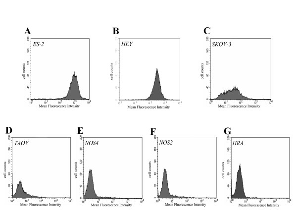
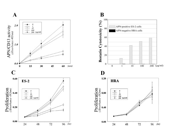
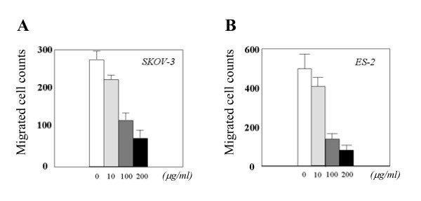
Methods: We first examined the expression of APN/CD13 at the protein level in a variety of OVCA cell lines and tissues. We subsequently investigated whether there was a correlation between APN/CD13 expression and invasive potential of various OVCA cell lines. Moreover, we investigated the function of APN/CD13 in OVCA cells using bestatin, an APN/CD13 inhibitor, or transfection of siRNA for APN/CD13.
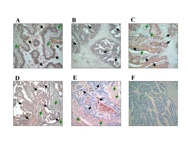
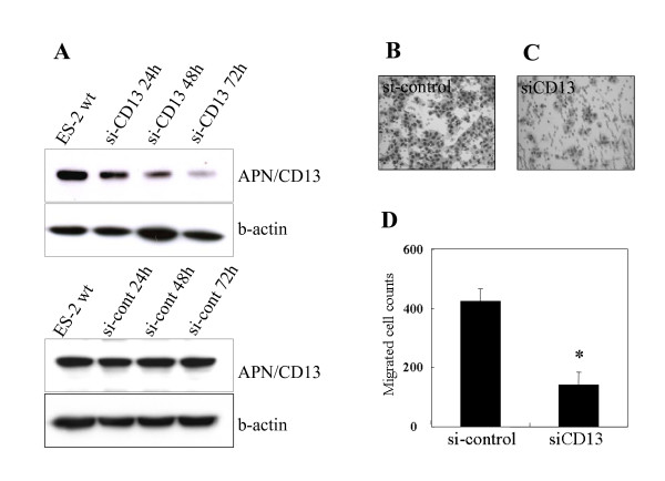
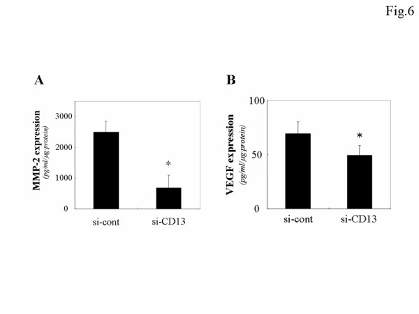
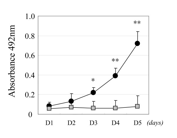
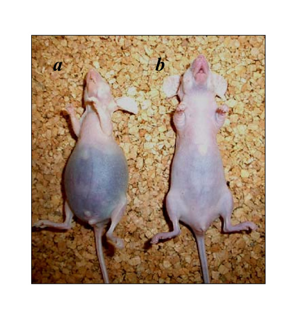
Results: We confirmed that APN/CD13 was expressed in OVCA tissues and cell lines to various extents. There was a positive correlation between APN/CD13 expression and migratory potential in various OVCA cell lines with accordingly enhanced secretion of endogenous MMP-2. Subsequently, we found a significant decrease in the proliferative and migratory abilities of OVCA cells after the addition of bestatin or the inhibition of APN/CD13 expression by siRNA. Furthermore, in an animal model, daily intraperitoneal administration of bestatin after inoculation of OVCA cells resulted in a decrease of peritoneal dissemination and in prolonged survival of nude mice.
Conclusion: The current data indicate the possible involvement of APN/CD13 in the development of OVCA, and suggest that clinical use of bestatin may contribute to better prognosis for ovarian carcinoma patients.
Figures
Similar articles
-
Involvement of aminopeptidase N in enhanced chemosensitivity to paclitaxel in ovarian carcinoma in vitro and in vivo.Int J Cancer. 2007 May 15;120(10):2243-50. doi: 10.1002/ijc.22528. Int J Cancer. 2007. PMID: 17266036
-
Targeting aminopeptidase N (APN/CD13) with cyclic-imide peptidomimetics derivative CIP-13F inhibits the growth of human ovarian carcinoma cells.Cancer Lett. 2010 Jun 28;292(2):153-62. doi: 10.1016/j.canlet.2009.11.021. Epub 2009 Dec 29. Cancer Lett. 2010. PMID: 20042271
-
Biological significance of aminopeptidase N/CD13 in thyroid carcinomas.Cancer Res. 2003 Dec 1;63(23):8500-6. Cancer Res. 2003. PMID: 14679016
-
Role of alanyl aminopeptidase in growth and function of human T cells (review).Int J Mol Med. 1999 Jul;4(1):17-27. Int J Mol Med. 1999. PMID: 10373632 Review.
-
Dual inhibition of dipeptidyl peptidase IV and aminopeptidase N suppresses inflammatory immune responses.Ann N Y Acad Sci. 2007 Sep;1110:402-9. doi: 10.1196/annals.1423.042. Ann N Y Acad Sci. 2007. PMID: 17911455 Review.
Cited by
-
Prognostic significance of aberrantly silenced ANPEP expression in prostate cancer.Br J Cancer. 2013 Feb 5;108(2):420-8. doi: 10.1038/bjc.2012.549. Epub 2013 Jan 15. Br J Cancer. 2013. PMID: 23322201 Free PMC article.
-
Near-Infrared Fluorescent Probes for the Detection of Cancer-Associated Proteases.ACS Chem Biol. 2021 Aug 20;16(8):1304-1317. doi: 10.1021/acschembio.1c00223. Epub 2021 Jul 27. ACS Chem Biol. 2021. PMID: 34315210 Free PMC article. Review.
-
Activity screening and structure-activity relationship of the hit compounds targeting APN/CD13.Fundam Clin Pharmacol. 2011 Apr;25(2):217-28. doi: 10.1111/j.1472-8206.2010.00844.x. Fundam Clin Pharmacol. 2011. PMID: 20636366 Free PMC article.
-
Prognostic value of matrix metalloproteinase-2 (MMP-2), matrix metalloproteinase-9 (MMP-9) and aminopeptidase N/CD13 in breast cancer patients.Med Oncol. 2012 Jun;29(2):561-9. doi: 10.1007/s12032-011-9984-y. Epub 2011 May 25. Med Oncol. 2012. PMID: 21611838
-
Advancement of fluorescent aminopeptidase probes for rapid cancer detection-current uses and neurosurgical applications.Front Surg. 2024 Mar 7;11:1298709. doi: 10.3389/fsurg.2024.1298709. eCollection 2024. Front Surg. 2024. PMID: 38516394 Free PMC article. Review.
References
-
- Kehlen A, Gohring B, Langner J, Riemann D. Regulation of the expression of aminopeptidase A, aminopeptidase N/CD13 and dipeptidylpeptidase IV/CD26 in renal carcinoma cells and renal tubular epithelial cells by cytokines and cAMP-increasing mediators. Clin Exp Immunol. 1998;111:435–441. doi: 10.1046/j.1365-2249.1998.00513.x. - DOI - PMC - PubMed
-
- Shipp MA, Look AT. Hematopoietic differentiation antigens that are membrane-associated enzymes: cutting is the key! Blood. 1993;82:1052–1070. - PubMed
MeSH terms
Substances
LinkOut - more resources
Full Text Sources
Other Literature Sources
Medical
Miscellaneous
