. 2007 Nov 10;368(1):91-101.
doi: 10.1016/j.virol.2007.06.023. Epub 2007 Jul 25.

Airway epithelial cell response to human metapneumovirus infection

Affiliations

Airway epithelial cell response to human metapneumovirus infection

X Bao et al. Virology. .

Abstract

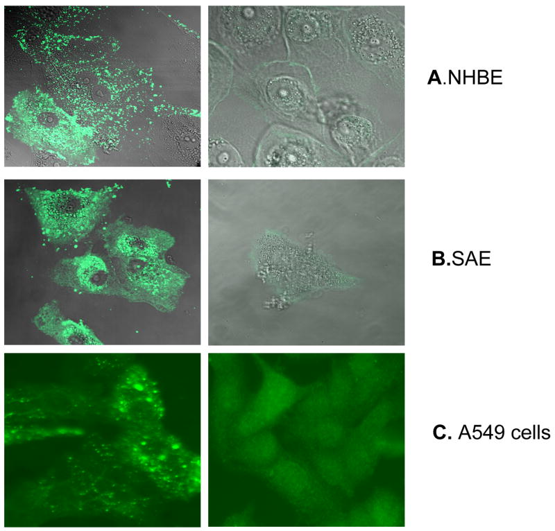
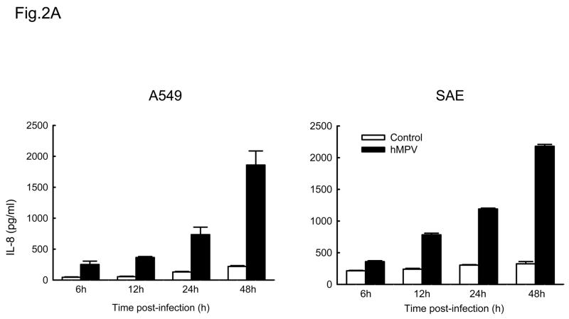
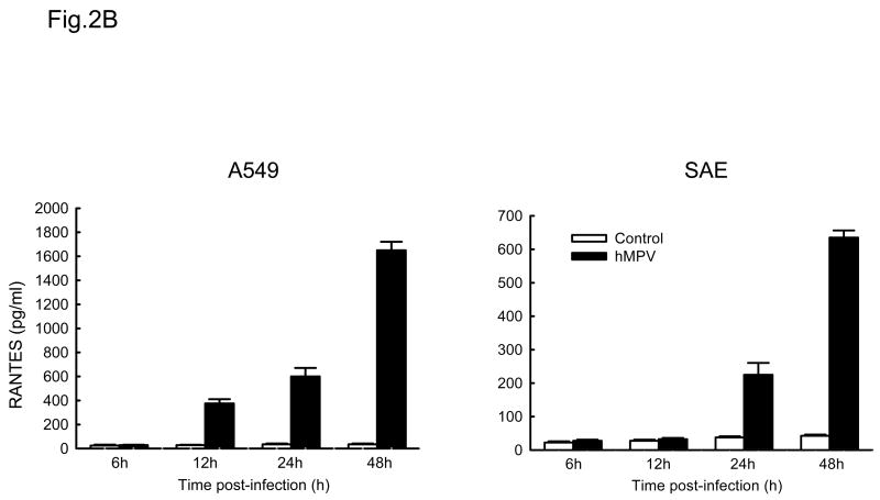
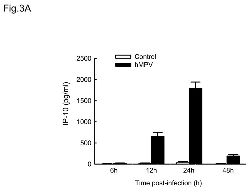
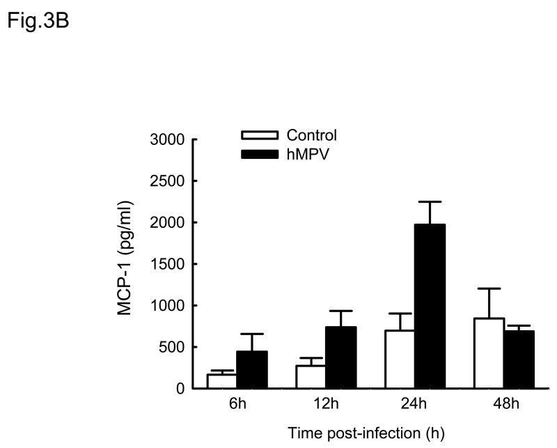
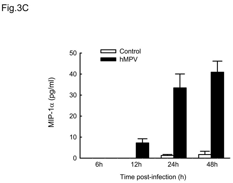
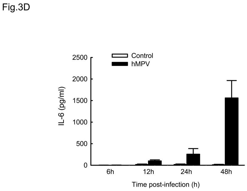
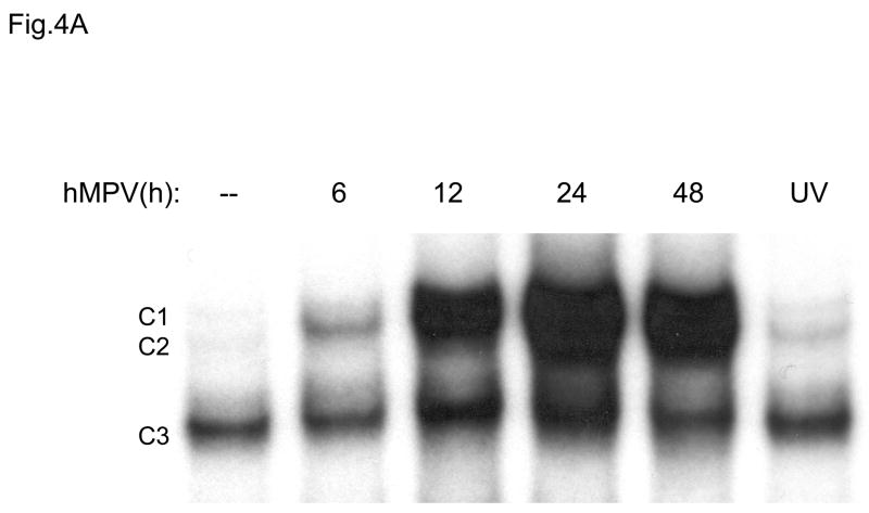
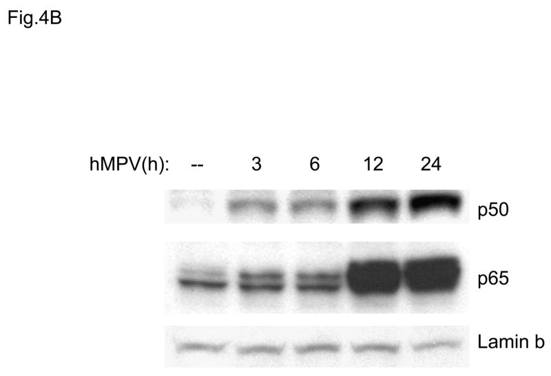
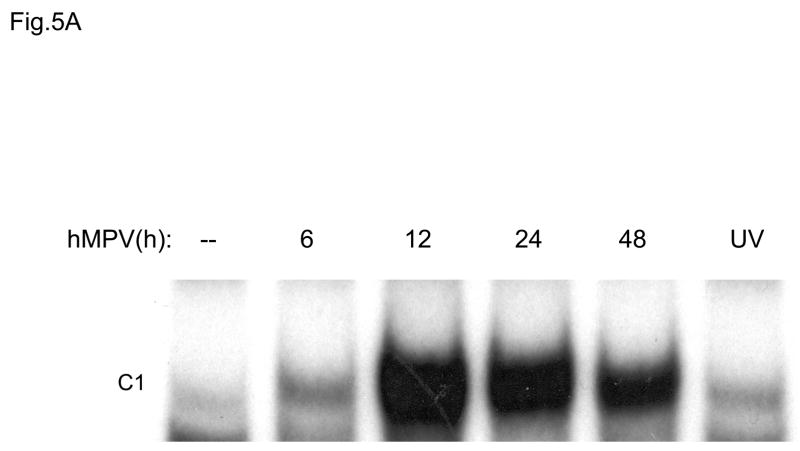
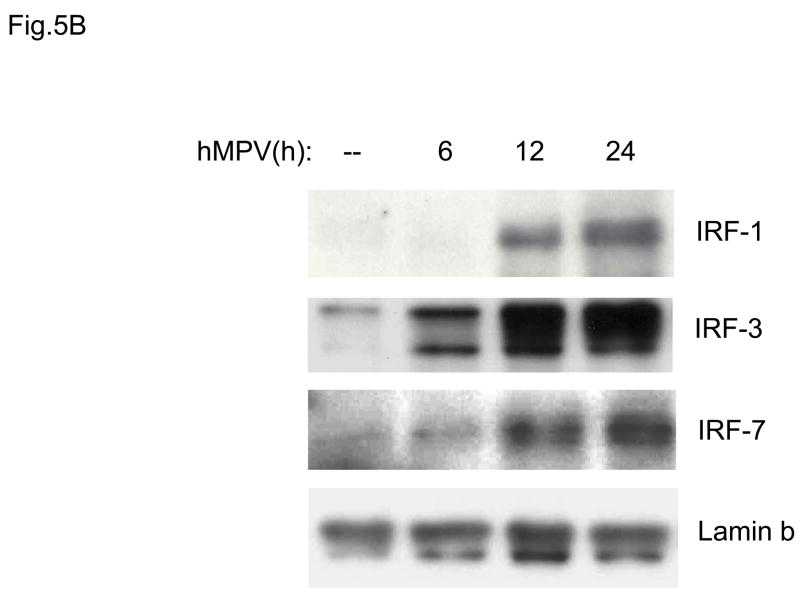
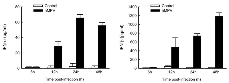
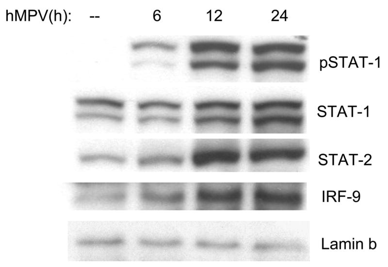
Human metapneumovirus (hMPV) is a major cause of lower respiratory tract infections (LRTIs) in infants, elderly and immunocompromised patients. In this study, we show that hMPV can infect in a similar manner epithelial cells representative of different tracts of the airways. hMPV-induced expression of chemokines IL-8 and RANTES in primary small alveolar epithelial cells (SAE) and in a human alveolar type II-like epithelial cell line (A549) was similar, suggesting that A549 cells can be used as a model to study lower airway epithelial cell responses to hMPV infection. A549 secreted a variety of CXC and CC chemokines, cytokines and type I interferons, following hMPV infection. hMPV was also a strong inducer of transcription factors belonging to nuclear factor (NF)-kappaB, interferon regulatory factors (IRFs) and signal transducers and activators of transcription (STATs) families, which are known to orchestrate the expression of inflammatory and immunomodulatory mediators.


Figures

Fig. 1
Fig. 1. Immunofluorescence of hMPV-infected human airway epithelial cells
Normal bronchial epithelial cells (NHBE, A), small airway epithelial cell (SAE, B) and human alveolar type II-like epithelial cell line (A549, C) were infected with hMPV at MOI 3 for 48 h and stained with nti-hMPV polyclonal antibody (left panel) or isotype antibody (right panel). Cells were analyzed using a Zeiss LSM 510 meta-ultraviolet laser scan confocal microscope for A and B or fluorescent microcopy for C. No staining was observed in mock-infected cells when similar staining was done (data not shown).
Fig. 2
Fig. 2. Chemokines induced by hMPV in A549 and SAE cells
A549 and SAE cells were infected with hMPV at MOI of 3 for various length of time. Secretion of IL-8 (A) and RANTES (B) by hMPV-infected A549 and SAE cells was assayed by ELISA. Data are expressed as mean ± standard error of two independent experiments performed in triplicates.
Fig. 3
Fig. 3. Cytokine and chemokine production by A549 cells infected with hMPV
A549 cells were infected with hMPV at MOI of 3. Cell supernatants were collected at different time points of infection and tested for cytokines and chemokine secretion by multiplex bead assay. Data are expressed as mean ± standard error of two independent experiments performed in triplicates. Open bars represent uninfected and solid bars hMPV-infected cells.
Fig. 4
Fig. 4. NF-kB activation in response to hMPV infection in A549 cells
(A). Nuclear extracts were prepared from A549 cells control and infected with hMPV, MOI of 3, for 6, 12, 24 and 48 h and used for binding to the IL-8 NF-kB probe in EMSA. Shown are the nucleoprotein complexes formed on the probe in control cells and in response to the infection. C1 and C2 are hMPV-inducible complexes, while C3 is constitutive. UV indicates UV-inactivated virus. (B) Western blot of p50 and p65 in hMPV-infected cells. Nuclear proteins were prepared from control and A549 cells infected for various length of time, fractionated on a 10% SDS-PAGE, transferred to PVDF membranes and probed with the appropriate antibody. Lamin b was used as an internal control to determine equal loading of the samples.
Fig. 5
Fig. 5. IRF activation induced by hMPV infection in A549 cells
(A). Nuclear extracts were prepared from A549 cells control and infected with hMPV, MOI of 3, for 6, 12, 24 and 48 h and used for binding to the RANTES ISRE probe in EMSA. Shown is the inducible nucleoprotein complex formed on the probe in response to the infection. UV indicates UV-inactivated virus. (B) Western blot of IRF-1,-3, and -7 in A549 cells infected with hMPV. Nuclear proteins were prepared from control and A549 cells infected for various length of time, fractionated on a 10% SDS-PAGE, transferred to PVDF membranes and probed with the appropriate antibody. Lamin b was used as an internal control to determine equal loading of the samples.
Fig. 6
Fig. 6. Type I IFN production by airway epithelial cells infected with hMPV
A549 cells were infected with hMPV at MOI of 3. Cell supernatants were collected at 6, 12, 24 and 48 h p.i. and tested for type I interferon production by ELISA. Data are expressed as mean ± standard error of two independent experiments performed in triplicates. Open bars represent uninfected and solid bars hMPV-infected cells.
Fig. 7
Fig. 7. ISGF3 complex activation in response to hMPV infection
A549 cells were infected with hMPV, MOI of 3, for 6, 12 and 24 h. Cells were harvested to prepare nuclear extracts and equal amounts of protein from control and infected cells were assayed for phosphorylated and total STAT1, total STAT2 and IRF-9 by Western blot. Lamin b was used as internal control to determine equal loading of the samples.

References

    1. Alvarez R, Harrod KS, Shieh WJ, Zaki S, Tripp RA. Human metapneumovirus persists in BALB/c mice despite the presence of neutralizing antibodies. J Virol. 2004;78:14003–14011. - PMC - PubMed
    1. Baggiolini M, Dewald B, Moser B. Human chemokines: an update. Annual Review Immunology. 1997;15:675–705. - PubMed
    1. Barnes B, Lubyova B, Pitha PM. On the role of IRF in host defense. J Interferon Cytokine Res. 2002;22:59–71. - PubMed
    1. Beg AA, Baldwin ASJ. The I kappa B proteins: multifunctional regulators of Rel/NF- kappa B transcription factors. Genes and Development. 1993;7:2064–2070. - PubMed
    1. Biacchesi S, Pham QN, Skiadopoulos MH, Murphy BR, Collins PL, Buchholz UJ. Modification of the trypsin-dependent cleavage activation site of the human metapneumovirus fusion protein to be trypsin independent does not increase replication or spread in rodents or nonhuman primates. J Virol. 2006;80:5798–5806. - PMC - PubMed

Publication types

MeSH terms