Comparative Study
. 2007 Aug 10;130(3):470-83.
doi: 10.1016/j.cell.2007.06.011. Epub 2007 Jul 26.

Sox17 dependence distinguishes the transcriptional regulation of fetal from adult hematopoietic stem cells

Affiliations
Comparative Study

Sox17 dependence distinguishes the transcriptional regulation of fetal from adult hematopoietic stem cells

Injune Kim et al. Cell. .

Abstract

Fetal stem cells differ phenotypically and functionally from adult stem cells in diverse tissues. However, little is known about how these differences are regulated. To address this we compared the gene expression profiles of fetal versus adult hematopoietic stem cells (HSCs) and discovered that the Sox17 transcriptional regulator is specifically expressed in fetal and neonatal but not adult HSCs. Germline deletion of Sox17 led to severe fetal hematopoietic defects, including a lack of detectable definitive HSCs. Conditional deletion of Sox17 from hematopoietic cells led to the loss of fetal and neonatal but not adult HSCs. HSCs stopped expressing Sox17 approximately 4 weeks after birth. During this transition, loss of Sox17 expression correlated with slower proliferation and the acquisition of an adult phenotype by individual HSCs. Sox17 is thus required for the maintenance of fetal and neonatal HSCs and distinguishes their transcriptional regulation from adult HSCs.


Figures
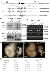
Figure 1
Figure 1. Generation of Sox17GFP/GFP Mice
(A) Schematic representation of the Sox17 targeted allele. The genomic structure after neo-cassette removal is shown on the fourth line. B and R indicate BamHI and EcoRV digestion sites, respectively. (B) Genotypes of heterozygous F1 mice (before neo excision) and F2 mice (after neo excision) from two independent lines were confirmed by Southern blot. Tail genomic DNA was digested with EcoRV and BamHI for 5′ and 3′ probe hybridization, respectively. (C) The lack of Sox17 expression in Sox17GFP/GFP embryos from both lines of mice was confirmed by RT-PCR using primers that amplified the Sox17 5′ untranslated region (which was expressed from the targeted allele), the coding sequence (not expressed), and the Sox17 3′ untranslated region (not expressed). (D) At E11.5, Sox17GFP/GFP embryos were much smaller than Sox17GFP/+ embryos and exhibited severe defects in posterior patterning (arrow) and body axis rotation (arrowheads). (E) Genotypes of progeny from Sox17GFP/+ intercrosses revealed Mendelian inheritance up to E12.5 but loss of Sox17-deficienct embryos by E13.5.
Figure 2
Figure 2. Sox17 Is Required for Hematopoiesis in the Fetal Liver and Yolk Sac
The yolk sac (A) and fetal liver (B) of E11.5 embryos contained obvious blood cells in Sox17GFP/+ embryos but not in Sox17GFP/GFP littermates. The numbers of CD45+ hematopoietic cells and Ter119+ erythroid cells in E11.5 yolk sac and whole embryo were significantly (*, p < 0.05) reduced in Sox17GFP/GFP embryos as compared to Sox17GFP/+ and Sox17+/+ embryos (C, three independent experiments). The numbers of colony-forming progenitors (CFU-C) in E11.5 yolk sac and whole embryo were also significantly (#, p < 0.01) reduced in Sox17GFP/GFP embryos as compared to Sox17GFP/+ and Sox17+/+ embryos (D, three independent experiments).
Figure 3
Figure 3. Sox17 Is Expressed by, and Required for the Maintenance of, Fetal and Neonatal But Not Adult HSCs
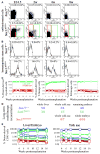
(A) Sox17 expression in fetal liver or bone marrow cells was analyzed by flow cytometry in Sox17GFP/+ mice (five independent experiments). Cells from Sox17+/+ mice established background. Less than 1% of fetal liver and neonatal bone marrow cells expressed Sox17 (green boxes). (B) HSCs were isolated from fetal liver as Sca-1+lineageMac-1+CD48 cells and from bone marrow as Sca-1+lineagec-kit+CD48 cells. Most fetal liver and neonatal bone marrow HSCs expressed Sox17, but expression began to decline by 4 weeks of age and was no longer evident at 8 weeks of age (five independent experiments). (C) Irradiated mice were transplanted with 1,000 GFP+ fetal liver cells (green lines) or 199,000 GFP fetal liver cells (red lines) from E14.5 Sox17GFP/+ donor mice. All mice transplanted with GFP+ cells were long-term multilineage reconstituted by donor cells (n = 12; each line represents a single mouse) but none of the mice transplanted with GFP cells showed detectable reconstitution (n = 11 mice; the black line at 0.3% represents background). (D) Irradiated recipient mice were transplanted with unfractionated fetal liver, yolk sac, or remaining embryo cells from single E12.5 Sox17+/+, Sox17GFP/+, or Sox17GFP/GFP embryos. Numbers indicate the fraction of recipient mice that were long-term multilineage reconstituted (>16 weeks) by donor cells. (E) While control cells usually gave long-term multilineage reconstitution (blue and green lines), we never detected any reconstitution by Sox17GFP/GFP cells (red lines).
Figure 4
Figure 4. Conditional Deletion of Sox17 in Hematopoietic/Endothelial Cells Leads to Severe Hematopoietic Defects and Lethality by E13.5
(A) Targeting of Sox17 to generate a floxed allele. Arrows indicate the sites in the wild-type allele (Sox17+) where loxP elements were inserted. These sites were selected to avoid disrupting conserved sequences (potential regulatory elements). Note that Cre-mediated recombination removes the entire Sox17 coding sequence. (B)The genotypes of heterozygous F1(Sox17fl-neo) and F2 (Sox17fl) mice from two independent lines were verified by Southern blot. Genomic tail DNA was digested with EcoRV (R) and SalI (Sl) for the 5′ probe and with SacI (S) for the 3′ probe. (C) The lack of Sox17 expression in CD144+ endothelial cells sorted from E10.5 Tie2-Cre+Sox17fl/GFP embryos was confirmed by RT-PCR using primers that amplified the Sox17 5′ untranslated region (expressed from the targeted allele), the coding sequence (not expressed) and the Sox17 3′ untranslated region (not expressed). (D) E12.5 Tie2-Cre+Sox17fl/GFP embryos were pale and growth retarded and lacked visible hematopoiesis. (E) Unlike control embryos, the yolk sac from E12.5 Tie2-Cre+Sox17fl/GFP embryos lacked visible hematopoiesis. (F) Progeny derived from mating Tie2-Cre+Sox17GFP/+ males with Sox17fl/fl females: conditional deletion of Sox17 using Tie2-Cre was lethal by E13.5.
Figure 5
Figure 5. Conditional Deletion of Sox17 Using Tie2-Cre Leads to a Failure to Generate or Maintain HSCs
The numbers of CD45+ or Ter119+ cells (A) and colony-forming progenitors (CFU-C; B) were dramatically reduced in the yolk sac and embryo of E11.5 Tie2-Cre+Sox17fl/GFP mice as compared to littermate controls (*, p < 0.05; #, p < 0.01; four independent experiments). (C) Irradiated wild-type mice were transplanted with unfractionated fetal liver, yolk sac, or remaining embryo cells from single E12.5 Tie2-Cre+Sox17fl/GFP embryos or littermate controls. The livers of Tie2-Cre+Sox17fl/GFP embryos were so hypocellular and pale that the entire embryos were dissociated and transplanted, rather than just fetal liver. Numbers indicate the fraction of recipient mice that were long-term multilineage reconstituted by donor cells. (D) While control cells usually gave long-term multilineage reconstitution (blue and green lines), we never detected any reconstitution above background (0.3%) from Tie2-Cre+Sox17fl/GFP cells (red lines). (E) CFU-GEMM colonies from E11.5 Sox17fl/GFP and Tie2-Cre+Sox17fl/GFP yolk sac were similar in size and appearance. (F) Sox17 deletion also did not affect the proportions of colony types, though the absolute number of all colonies was reduced (three independent experiments). (G) E11.5 Tie2-Cre+Sox17fl/GFP and Sox17fl/GFP yolk sac cells expressed embryonic (β-H1) and adult (β-major) hemoglobin in vivo. In culture, all colonies from Sox17fl/GFP yolk sac (19/19) and Tie2-Cre+Sox17fl/GFP yolk sac (20/20) expressed adult hemoglobin (β-major). PCR on genomic DNA from individual colonies demonstrated that 95% of Tie2-Cre+Sox17fl/GFP colonies (19/20) had completely recombined Sox17 (data not shown).
Figure 6
Figure 6. Sox17 Is Autonomously Required for the Maintenance of Neonatal But Not Adult HSCs
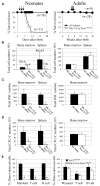
(A) Mx-1-Cre+Sox17fl/GFP mice and littermate controls were administered pIpC 2, 4, and 6 days after birth (arrows). All Mx-1-Cre+ Sox17fl/GFP mice died by 14 days after birth, but no littermate controls died. Adult Mx-1-Cre+Sox17fl/GFP mice that were administered seven doses of pIpC over a 14 day period beginning 6 weeks after birth all survived. (B–C) Four to five days after ending pIpC treatment in neonates and seven days after ending pIpC treatment in adults, bone marrow cellularity in the tibias and femurs and spleen cellularity were significantly (*, p < 0.05) reduced in neonatal Mx-1-Cre+Sox17fl/GFP mice (n = 5–7) as compared to littermate controls (n = 4–6) but were not affected in adult Mx-1-Cre+Sox17fl/GFP mice (n = 4–5) as compared to littermate controls (n = 4–5). The absolute numbers of Flk2Sca-1+Lineagec-kit+CD48 HSCs (C) and colony-forming progenitors (CFU-C; D) in the bone marrow (from tibias and femurs) and spleen were also significantly (p < 0.05) reduced in the same neonatal Mx-1-Cre+Sox17fl/GFP mice but were not affected in the adult Mx-1-Cre+Sox17fl/GFP mice. (E) Bone marrow cells from pIpC-treated neonatal Mx-1-Cre+Sox17fl/GFP mice (CD45.2+) failed to give long-term multilineage reconstitution in irradiated CD45.1+ wild-type recipients (n = 5), in contrast to the same dose of cells from littermates or adult mice (n = 4 or 5; differences in myeloid, B, and T cell chimerism were highly statistically significant: #, p < 0.001; 6 week time point is shown). In each case, 200,000 donor bone marrow cells were competed against 200,000 recipient bone marrow cells.
Figure 7
Figure 7. Sox17 Deletion from Neonatal HSCs Induces Cell Death, and the Postnatal Decline in Sox17 Expression by Wild-Type HSCs Is Associated with a Transition to an Adult Phenotype
(A and B) Sca-1+Lineagec-kit+CD48 HSCs isolated from the bone marrow of neonatal Mx-1-Cre+Sox17fl/GFP mice 5 days after pIpC treatment exhibited normal cell-cycle distribution relative to littermate controls (A) but a 3-fold increased frequency of cells undergoing cell death (Annexin V+; B). (C–F) GFP+ (Sox17-expressing) and GFP (Sox17-non-expressing) Sca-1+Lineagec-kit+CD48 HSCs were isolated from the bone marrow of 3.5 to 4 week-old Sox17GFP/+ mice. GFP Sca-1+Lineagec-kit+CD48 HSCs were dividing less rapidly (D), and failed to express AA4.1 (E) or Mac-1 (F), consistent with an adult HSC phenotype. In contrast, GFP+ Sca-1+Lineagec-kit+CD48 HSCs were more rapidly dividing (D), and expressed AA4.1 (E) and Mac-1 (F), consistent with a fetal HSC phenotype.

Comment in

Similar articles

Cited by

References

    1. Azcoitia V, Aracil M, Martinez AC, Torres M. The homeodomain protein Meis1 is essential for definitive hematopoiesis and vascular patterning in the mouse embryo. Dev Biol. 2005;280:307–320. - PubMed
    1. Bowie MB, McKnight KD, Kent DG, McCaffrey L, Hoodless PA, Eaves CJ. Hematopoietic stem cells proliferate until after birth and show a reversible phase-specific engraftment defect. J Clin Invest. 2006;116:2808–2816. - PMC - PubMed
    1. Chambers I, Colby D, Robertson M, Nichols J, Lee S, Tweedie S, Smith A. Functional expression cloning of Nanog, a pluripotency sustaining factor in embryonic stem cells. Cell. 2003;113:643–655. - PubMed
    1. Cobas M, Wilson A, Ernst B, Mancini SJ, MacDonald HR, Kemler R, Radtke F. {beta}-Catenin Is Dispensable for Hematopoiesis and Lymphopoiesis. J Exp Med. 2004;199:221–229. - PMC - PubMed
    1. Davidson AJ, Ernst P, Wang Y, Dekens MP, Kingsley PD, Palis J, Korsmeyer SJ, Daley GQ, Zon LI. cdx4 mutants fail to specify blood progenitors and can be rescued by multiple hox genes. Nature. 2003;425:300–306. - PubMed

Publication types

MeSH terms