. 2007 Oct 15;159(1):85-101.
doi: 10.1016/j.resp.2007.06.002. Epub 2007 Jun 16.

Arrest of 5HT neuron differentiation delays respiratory maturation and impairs neonatal homeostatic responses to environmental challenges

Affiliations

Arrest of 5HT neuron differentiation delays respiratory maturation and impairs neonatal homeostatic responses to environmental challenges

Jeffery T Erickson et al. Respir Physiol Neurobiol. .

Abstract

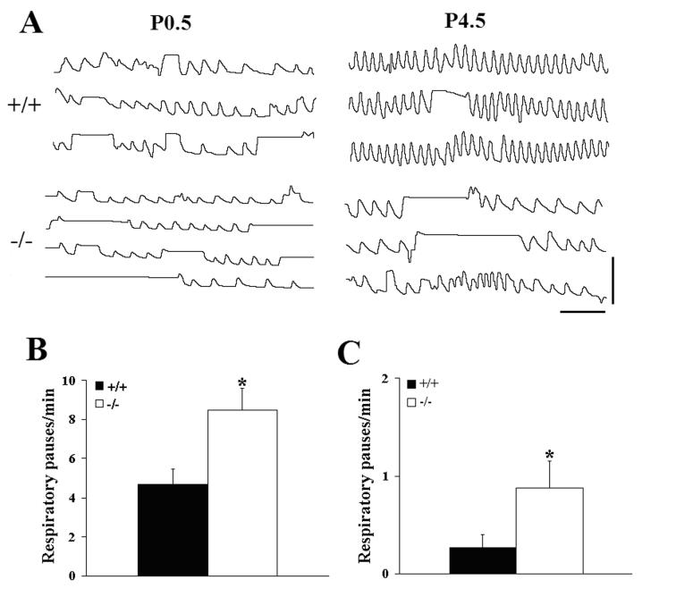
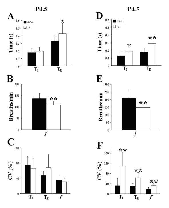
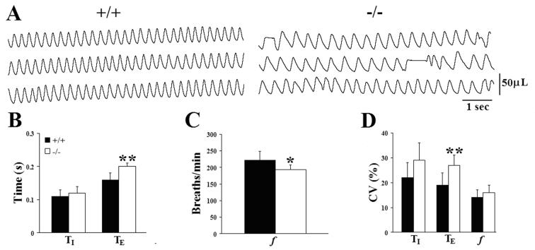
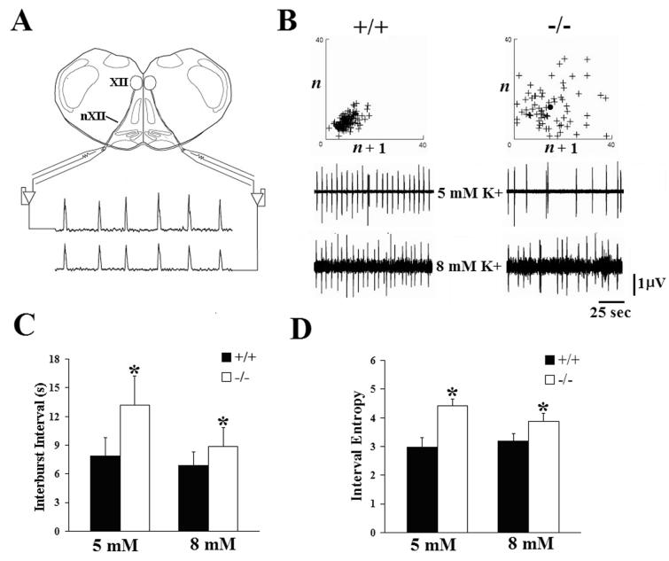
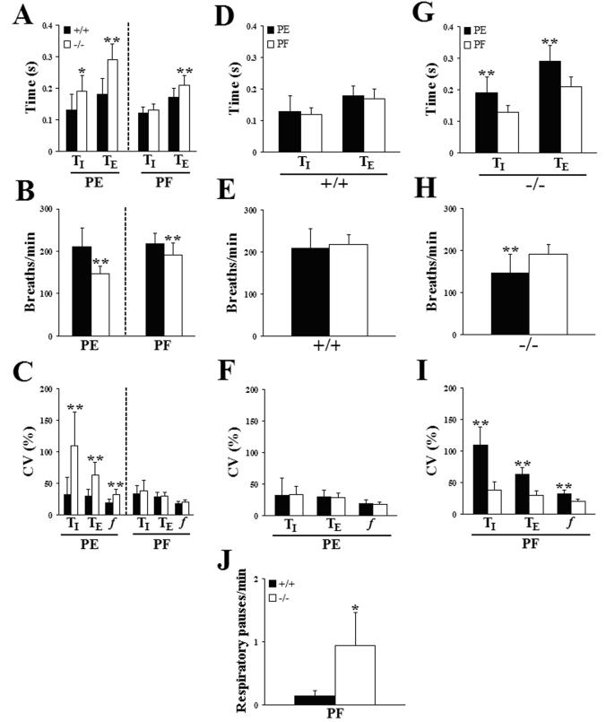
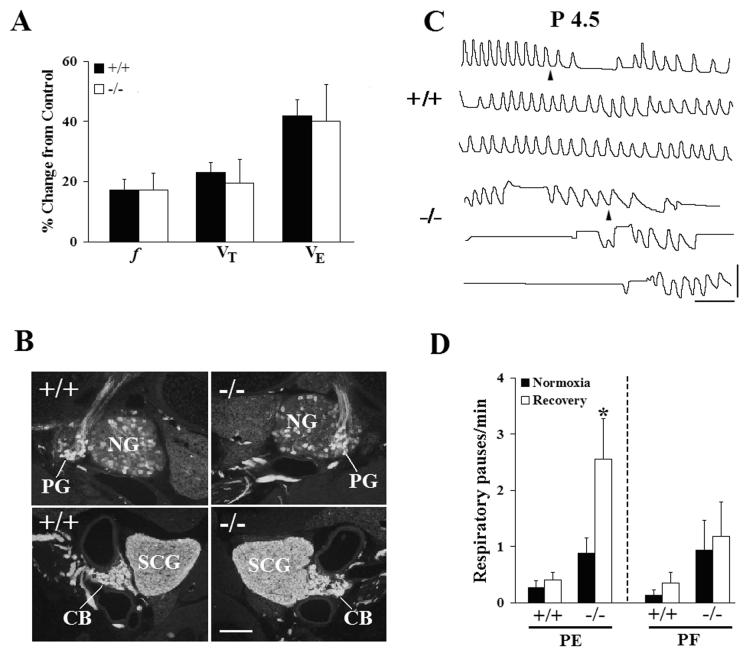
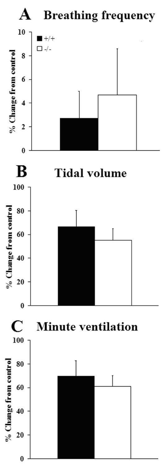
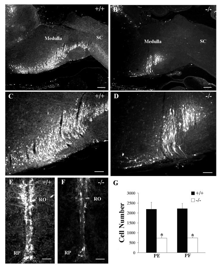
Serotonin (5HT) is a powerful modulator of respiratory circuitry in vitro but its role in the development of breathing behavior in vivo is poorly understood. Here we show, using 5HT neuron-deficient Pet-1 (Pet-1(-/-)) neonates, that serotonergic function is required for the normal timing of postnatal respiratory maturation. Plethysmographic recordings reveal that Pet-1(-/-) mice are born with a depressed breathing frequency and a higher incidence of spontaneous and prolonged respiratory pauses relative to wild type littermates. The wild type breathing pattern stabilizes by postnatal day 4.5, while breathing remains depressed, highly irregular and interrupted more frequently by respiratory pauses in Pet-1(-/-) mice. Analysis of in vitro hypoglossal nerve discharge indicates that instabilities in the central respiratory rhythm generator contribute to the abnormal Pet-1(-/-) breathing behavior. In addition, the breathing pattern in Pet-1(-/-) neonates is susceptible to environmental conditions, and can be further destabilized by brief exposure to hypoxia. By postnatal day 9.5, however, breathing frequency in Pet-1(-/-) animals is only slightly depressed compared to wild type, and prolonged respiratory pauses are rare, indicating that the abnormalities seen earlier in the Pet-1(-/-) mice are transient. Our findings provide unexpected insight into the development of breathing behavior by demonstrating that defects in 5HT neuron development can extend and exacerbate the period of breathing instability that occurs immediately after birth during which respiratory homeostasis is vulnerable to environmental challenges.


Figures

Figure 1
Figure 1
Breathing behavior is compromised in Pet-1−/− mice during the early postnatal period. (A) Plethysmographic recordings of resting ventilation in wild-type (+/+) and Pet-1 null (−/−) mice made within 12 hrs of birth (P0.5) and on postnatal day 4.5 (P4.5) while breathing 21% O2. The −/− recordings reflect the finding that the prolonged respiratory pauses occurred predominantly during inspiration. (B,C) Comparison of the incidence of prolonged (≥1 sec duration) respiratory pauses in wild type (+/+) and Pet-1 mutant (−/−) mice on the day of birth (B; +/+, 4.7 ± 0.78/min, n = 13 vs −/−, 8.5 ± 4.1/min, n = 13, P=0.0114) and on postnatal day 4.5 (C; +/+, 0.27 ± 0.13/min, n = 19 vs −/−, 0.88 ± 0.27/min, n = 10, P=0.0288). Values are means ± SE. *P<0.05. Scale bars: horizontal, 2 sec; vertical, 50 μL.
Figure 2
Figure 2
Comparison of mean inspiratory (TI) and expiratory (TE) time, breathing frequency (f), and the coefficients of variation (CV=SD/mean × 100) of these parameters, respectively, on the day of birth (P0.5; A,B,C) and on postnatal day 4.5 (D,E,F). Values are means ± SD. *P<0.05; **P<0.01. See Table 2A,B for sample sizes and P values.
Figure 3
Figure 3
Breathing deficits improve in Pet-1−/− mice with increasing postnatal age. (A) Plethysmographic recordings of resting ventilation in a wild type (+/+) and Pet-1 mutant (−/−) mouse breathing 21% O2 on postnatal day 9.5. Recordings are from the same P4.5 animals presented in Fig. 1. (B,C,D) Comparison of mean inspiratory (TI) and expiratory (TE) time (B), breathing frequency (f; panel C), and the coefficients of variation (CV=SD/mean x 100) of these parameters (D) between genotypes on P9.5. At this age prolonged respiratory pauses were rarely seen in the serotonin-deficient mice. Values are means ± SD. *P<0.05; **P<0.01. See Table 2C for sample sizes and P values.
Figure 4
Figure 4
Disorder of central respiratory rhythm may contribute to the breathing deficits of Pet-1 null mutant mice. (A) In vitro recording arrangement. Output from the central respiratory rhythm generator was assessed via recordings from hypoglossal nerve (nXII) rootlets arising from neurons in the hypoglossal nucleus (XII). (B) XIIth nerve recordings from wild type (+/+, left panel) and Pet-1−/− (−/−, right panel) medullary slices at two different levels of extracellular potassium (5 and 8 mM K+). No significant differences in either burst duration or burst area were detected. Above each set of recordings are representative Poincaré plots (see Materials and Methods) of interburst intervals (analogous to TE in vivo) derived from recordings in +/+ and −/− slices at 8 mM K+. Each cross (+) represents a single nerve discharge. If interburst intervals were identical for every fictive breath, the graph would reduce to a single point representing that identical time between two discharges. (C) Comparison of mean interburst interval of XIIth nerve discharges from +/+ and −/− brainstem slices exposed to either 5 or 8 mM potassium. (D) Comparison of interval entropy values derived from XIIth nerve discharges in +/+ and −/−brainstem slices exposed to two different levels of extracellular potassium. For both panels (C) and (D): +/+: n=13 at 5 mM, n=19 at 8 mM; −/−: n=13 at 5 mM, n=15 at 8 mM. All values are means ± SD. *P<0.05.
Figure 5
Figure 5
The severity of breathing abnormalities in Pet-1−/− mice can be influenced by environmental conditions. (A,B,C) Comparison of inspiratory (TI) and expiratory (TE) time (A), breathing frequency (B) and the coefficients of variation of these ventilatory parameters (C) in P4.5 wild type (+/+, dark bars) and Pet-1 null mutant (−/−, light bars) mice reared in either a pathogen-exposed (PE) or pathogen-free (PF) environment. (D,E,F) Direct comparison of inspiratory (TI) and expiratory (TE) time (D), breathing frequency (E) and the coefficients of variation of these ventilatory parameters (F) in P4.5 +/+ mice from the PE and PF colonies. (G,H,I) Direct comparison of inspiratory (TI) and expiratory (TE) time (G), breathing frequency (H) and the coefficients of variation of these ventilatory parameters (I) in P4.5 Pet-1−/− mice from the PE and PF colonies. (J) Pet-1−/− mice from the PF colony also suffered an increased incidence of prolonged respiratory pauses, relative to wild type littermates (compare with Fig. 1C). Values are means ± SD. *P<0.05; **P<0.01. See Table 2B, Table 3 for sample sizes and P values.
Figure 6
Figure 6
Breathing behavior in Pet-1−/− mice can be destabilized following a short-term exposure to low environmental oxygen. (A) Pet-1−/− mice are able to detect a low oxygen stimulus. No differences in breathing frequency (f), tidal volume (VT) or minute ventilation (VE) were observed in P4.5 +/+ or −/− mice from the PE colony during the initial 15s of a 2 minute exposure to 10% O2. (B) Tyrosine hydroxylase (TH)-immunostained sections through the petrosal ganglion (upper panels) and carotid body (lower panels) from a +/+ and −/− mouse. (C) Comparison of plethysmographic recordings from a P4.5 wild type (+/+) and Pet-1 mutant (−/−) mouse during the transition (arrowhead) from 10% to 21% O2, following a 2 min hypoxic exposure. Several long respiratory pauses can be seen in the record from the mutant mouse immediately following the low O2 exposure. (D) Comparison of the incidence of long respiratory pauses in +/+ and −/− mice from either the pathogen-exposed (PE) or pathogen-free (PF) colony while breathing 21% O2 before (Normoxia) or during a 5 minute normoxic period following a 2 min exposure to 10% O2 (Recovery). A significant increase in the incidence of hypoxia-induced pauses was seen only in −/− mice from the PE colony. Values are means ± SE. Scale bars: panel B, 100 μm; panel C, horizontal, 2 sec, vertical, 50 μL. *P<0.05.
Figure 7
Figure 7
Breathing responses of P4.5 wild type (+/+, n=12) and Pet-1 null mutants (−/−, n=8) from the PF colony to 5% CO2 (in 21% O2, balance N2) expressed as mean percent change from pre-stimulus control levels. Measurements were made during the final minute of a two minute stimulation period. No significant differences (two-tailed t-test, α=0.05) were observed between genotypes in breathing frequency (A), tidal volume (B) or minute ventilation (C). Values are means ± SE.
Figure 8
Figure 8
Pet-1−/− mice reared in the PE and PF colonies have a similar degree of 5HT cell loss in the medulla oblongata. Photomicrographs of sagittal (A-D) or transverse (E,F) sections of the medulla oblongata of a wild type (A, 4x; C, 10x, E, 20x) and a Pet-1−/− (B, 4x; D, 10x, F, 20x) mouse from the PF colony that were immunostained to detect 5HT. (G) The total number of 5HT-immunostained cells was reduced by 67% in the null mutants from the PE colony and by 66% in mutants from the PF colony, relative to their respective wild type controls. Two-way ANOVA revealed significant differences between genotypes in each colony (treatment effect, F(1,16) = 219.2, P<0.0001), but no differences between genotypes across colonies (location effect, F(1,16) = 0.051, P=0.824) and no significant interaction effect (F(1,16) = 0.009, P=0.924). n = 5 for each group. RO, nucleus raphe obscurus; RP, nucleus raphe pallidus; SC, spinal cord. Scale bars: A,B = 250 μm; C,D = 100 μm; E,F = 50 μm.

References

    1. Abadie V, Champagnat J, Fortin G. Branchiomotor activities in mouse embryo. NeuroReport. 2000;11:141–145. - PubMed
    1. Allain AE, Meyrand P, Branchereau P. Ontogenetic changes of the spinal GABAergic cell population are controlled by the serotonin (5-HT) system: implication of the 5-HT1 receptor family. J. Neurosci. 2005;25:8714–8724. - PMC - PubMed
    1. Al-Zubaidy Z, Erickson R, Greer J. Serotonergic and noradrenergic effects on respiratory neural discharges in the medullary slice preparation of neonatal rats. Pflügers Arch. 1996;431:942–949. - PubMed
    1. Bartlett D, Remmers JE, Jr., Gautier H. Laryngeal regulation of respiratory airflow. Respir. Physiol. 1973;18:194–200. - PubMed
    1. Bou-Flores C, Lajard AM, Monteau R, De Maeyer E, Seif I, Lanoir J, Hilaire G. Abnormal phrenic motoneuron activity and morphology in neonatal monoamine oxidase A-deficient transgenic mice: possible role of serotonin excess. J. Neurosci. 2000;20:4646–4656. - PMC - PubMed

Publication types

LinkOut - more resources