. 2007 Sep;18(9):2509-16.
doi: 10.1681/ASN.2007030348. Epub 2007 Jul 26.

Thiazide diuretics directly induce osteoblast differentiation and mineralized nodule formation by interacting with a sodium chloride co-transporter in bone

Affiliations

Thiazide diuretics directly induce osteoblast differentiation and mineralized nodule formation by interacting with a sodium chloride co-transporter in bone

Melita M Dvorak et al. J Am Soc Nephrol. 2007 Sep.

Abstract

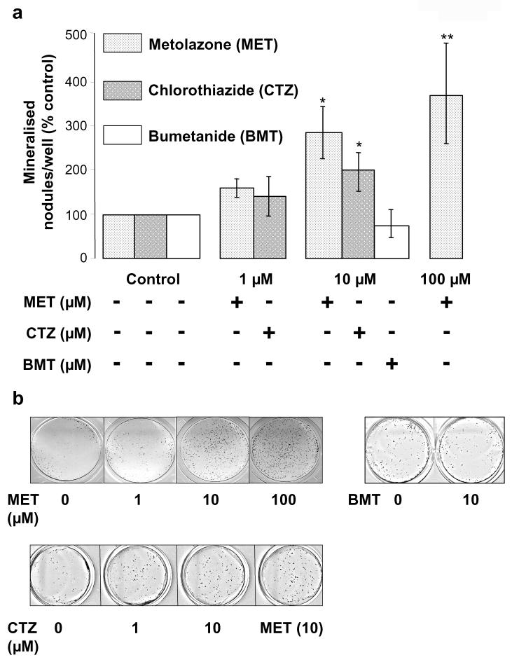
Thiazide diuretics are used worldwide as a first-choice drug for patients with uncomplicated hypertension. In addition to their antihypertensive effect, thiazides increase bone mineral density and reduce the prevalence of fractures. Traditionally, these effects have been attributed to increased renal calcium reabsorption that occurs secondary to the inhibition of the thiazide-sensitive sodium chloride cotransporter (NCC) in the distal tubule. The aim of the current study was to determine whether thiazides exert a direct bone-forming effect independent of their renal action. We found that the osteoblasts of human and rat bone also express NCC, suggesting that these bone-forming cells may be an additional target for thiazides. In vitro, NCC protein was virtually absent in proliferating human and fetal rat osteoblasts, whereas its expression dramatically increased during differentiation. Thiazides did not affect osteoblast proliferation, but directly stimulated the production of the osteoblast differentiation markers runt-related transcription factor 2 (runx2) and osteopontin. Using overexpression/knockdown studies in fetal rat calvarial cells, we show that thiazides increase the formation of mineralized nodules, but loop diuretics do not. Overall, our study demonstrates that thiazides directly stimulate osteoblast differentiation and bone mineral formation independent of their effects in the kidney. Therefore, in addition to their use as antihypertensive drugs, our results suggest that thiazides may find a role in the prevention and treatment of osteoporosis.


Figures

Figure 1
Figure 1. NCC is expressed in bone
Using human NCC antibodies, specific immunofluorescence is evident in undecalcified frozen, human (a) and rat (b) sections. NCC is expressed in reversal lines (a; arrows), osteocytes (a; small arrows) and lining osteoblasts (b; arrows). c: Bright field photomicrograph of EDTA-decalcified paraffin section of rat femur show NCC immunoperoxidase activity (brown pigment) in reversal lines (arrows), osteoblasts (arrowheads) and some osteocytes (arrowheads +). A proportion of osteocytes are negative for NCC (arrowheads -). (d): Omission of primary antibodies does not result in staining. (e-i): Photomicrographs of rat femur cryosections show that NCC immunofluorescence is in reversal lines (e, f; arrows), osteoblasts (g, arrows), osteocytes (h; arrows) and some osteoclasts (i; arrows). j: NCC-specific, distal convoluted tubule staining is present in the rat kidney. Bar = 20μm throughout. (k, upper panel): NCC immunoreactivity in osteoblast-derived MG63 cells (ob, 30 μg of total homogenate loaded) is comparable to that detected in human kidney cortex sample (kc, 30 μg), with predicted sizes for the NCC monomer (M) and dimer (D) of ∼160 and 320 kDa, respectively. (k, lower panel): expression of the ubiquitously expressed, type 1 Na+-K+-2Cl- cotransporter (NKCC1) is also expressed in MG63 cells using an anti-rat NKCC1 antibody (ob, 30 μg of total homogenate loaded). The figure also shows that, as expected, NKCC1 is low abundant in human kidney cortex and outer medulla. (kc = kidney cortex, 30 μg; om = kidney outer medulla, 10 μg).
Figure 2
Figure 2. NCC is expressed in differentiating osteoblasts
(a): Western analysis of protein homogenates extracted from established models of osteoblasts (human UMR-106, murine 2T3 and rat fetal rat calvaria, FRC) reveal that NCC immunoreactivity is of comparable size to that observed in rat kidney. (b): Enzymatic deglycosylation of protein extracts from the human osteoblast-derived cell line MG63 cells indicates a reduction in the NCC monomer molecular weight from ∼160 kDa to ∼120 kDa. Control protein exposed to 37°C for 60 min without N-glycanase showed presence of the fully glycosylated protein. (c, d): NCC protein expression is absent in pre-confluent, proliferating FRC (c) and MG63 cells (d). In both models, NCC expression increases as the differentiation process progresses and peaks around two weeks post-confluency. Note that 0 days post-confluence represents the beginning of differentiation.
Figure 3
Figure 3. Metolazone does not affect proliferation but stimulates the expression of osteoblast differentiation markers
(a): FRC cell proliferation (for up to two weeks in culture) is not affected by increasing doses of the thiazide-like metolazone. (b): Consistent with the lack of NCC expression at days 1-3 post-confluency (see Figures 2c and 2d), the activity of collagen 1A (coll1A), the major structural component of the organic matrix and early differentiation marker, is not affected by 48h treatment with metolazone (MET, 10 μM) or with chlorothiazide (CTZ, 10 μM). (c-f): Western analyses show increased expression of osteoblast markers Runx2 (c and d) and osteopontin (e and f) in rat (c and e) and human (d and f) osteoblast models in response to increasing concentrations of metolazone (7 day treatment). Histograms represent mean band densities and indicate that the levels of expression of Runx2 or osteopontin, normalized for the levels of expression of β-actin in the same samples, are significantly increased [3c: *: p<0.05 at 10 μM (ANOVA, Tukey post hoc test). *: 3d: p<0.01 (unpaired Student’s t-test). 3e: *: p<0.05 (ANOVA, Tukey post hoc test). 3f *: p<0.05 (unpaired Student’s t-test)].
Figure 4
Figure 4. Thiazides stimulate mineralized nodule formation by FRC cells
(a): Continuous treatment of FRC cells with metolazone (MET, 1-100 μM) and chlorothiazide (CTZ, 1 and 10 μM) for 10-21 days post-confluency induces dose-dependent increases in mineralized nodule formation (MET: n=3; *: p<0.05, **: p<0.01. CTZ: n=3; *: p<0.05). The loop diuretic bumetanide (BMT, 10 μM) does not significantly affect nodule formation. (b): Representative von Kossa staining of FRC cells (15 days post-confluency) treated with MET (1-100 μM) shows a concentration-dependent increase in the number of mineralized nodules (upper panel) while no effects on FRC cell mineralization are seen in the presence of BMT (10 μM). Lower panel: the number of mineralized nodules is comparable for metolazone and chlorothiazide treated post-confluent FRC cells (representative of 3 independent cell isolations).
Figure 5
Figure 5. Metolazone-induced mineralization is mediated by NCC
(a): Metolazone treatment evokes a significant increase in mineralization in post-confluent FRC cells transiently transfected with empty vector (pcDNA3.1. *: p<0.05, n=9 from 3 independent cell isolations). (b): Metolazone-dependent (10 μM) mineralization in post-confluent FRC cells is dramatically enhanced by NCC overexpression (S, Sense) and this is significantly blunted by NCC knockdown (AS, antisense; p<0.05; n=12 observations made from four independent cell isolations). Note that NCC overexpression only increases mineralization of post-confluent FRC cells after metolazone treatment. Data are expressed as change in number of nodules per well, defined as [(NodulesNCC - NodulespcDNA)/NodulespcDNA] where NodulespcDNA = number of mineralized nodules in cells transfected with empty vector and NodulesNCC = number of mineralized nodules in cells transfected with NCC sense or antisense, as indicated below bars. (c): representative von Kossa staining of FRC cells following the treatments indicated below each well.

Similar articles

Cited by

References

    1. Gamba G. Molecular physiology and pathophysiology of electroneutral cation-chloride cotransporters. Physiol Rev. 2005;85(2):423–493. - PubMed
    1. Wasnich RD, Benfante RJ, Yano K, Heilbrun L, Vogel JM. Thiazide effect on the mineral content of bone. N Engl J Med. 1983;309(6):344–347. - PubMed
    1. LaCroix AZ, Wienpahl J, White LR, Wallace RB, Scherr PA, George LK, Cornoni-Huntley J, Ostfeld AM. Thiazide diuretic agents and the incidence of hip fracture. N Engl J Med. 1990;322(5):286–290. - PubMed
    1. Jones G, Nguyen T, Sambrook PN, Eisman JA. Thiazide diuretics and fractures: can meta-analysis help? J Bone Miner Res. 1995;10(1):106–111. - PubMed
    1. Brickman AS, Massry SG, Coburn JW. Changes in serum and urinary calcium during treatment with hydrochlorothiazide: studies on mechanisms. J Clin Invest. 1972;51(4):945–54. - PMC - PubMed

Publication types