. 2007 Aug 7;104(32):13176-81.
doi: 10.1073/pnas.0704533104. Epub 2007 Jul 26.

Correlated network activity enhances synaptic efficacy via BDNF and the ERK pathway at immature CA3 CA1 connections in the hippocampus

Affiliations

Correlated network activity enhances synaptic efficacy via BDNF and the ERK pathway at immature CA3 CA1 connections in the hippocampus

Majid H Mohajerani et al. Proc Natl Acad Sci U S A. .

Abstract

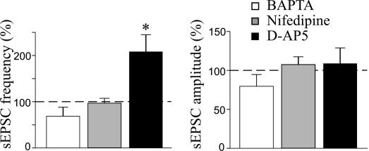
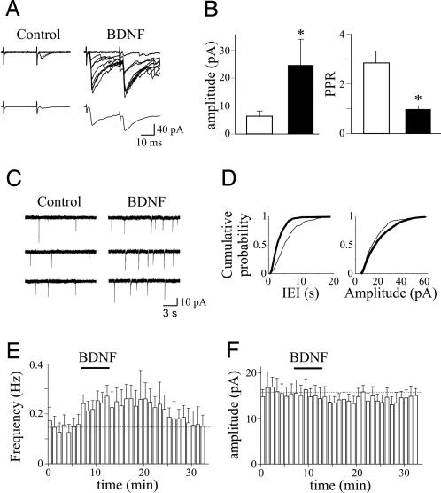
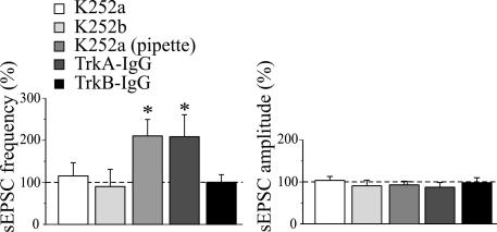
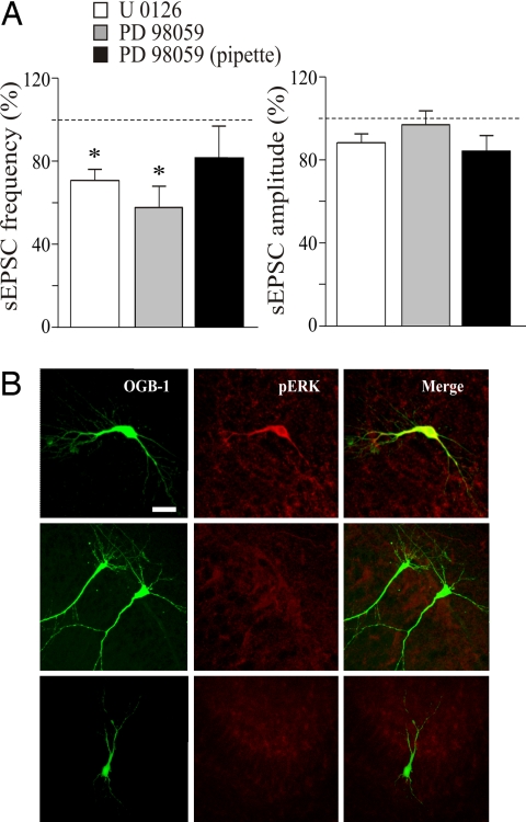
At early developmental stages, correlated neuronal activity is thought to exert a critical control on functional and structural refinement of synaptic connections. In the hippocampus, between postnatal day 2 (P2) and P6, network-driven giant depolarizing potentials (GDPs) are generated by the synergistic action of glutamate and GABA, which is depolarizing and excitatory. Here the rising phase of GDPs was used to trigger Schaffer collateral stimulation in such a way that synchronized network activity was coincident with presynaptic activation of afferent input. This procedure produced a persistent increase in spontaneous and evoked alpha-amino-3-hydroxy-5-methyl-4-isoxadepropionic acid-mediated glutamatergic currents, an effect that required calcium influx through postsynaptic L-type calcium channels. No potentiation was observed when a delay of 3 sec was introduced between GDPs and afferent stimulation. Pairing-induced potentiation was prevented by scavengers of endogenous BDNF or tropomyosin-related kinase receptor B (TrkB) receptor antagonists. Blocking TrkB receptors in the postsynaptic cell did not prevent the effects of pairing, suggesting that BDNF, possibly secreted from the postsynaptic cell during GDPs, acts on TrkB receptors localized on presynaptic neurons. Application of exogenous BDNF mimicked the effects of pairing on synaptic transmission. In addition, pairing-induced synaptic potentiation was blocked by ERK inhibitors, suggesting that BDNF activates the MAPK/ERK cascade, which may lead to transcriptional regulation and new protein synthesis in the postsynaptic neuron. These results support the hypothesis that, during a critical period of postnatal development, GABAA-mediated GDPs are instrumental in tuning excitatory synaptic connections and provide insights into the molecular mechanisms involved in this process.


Conflict of interest statement

The authors declare no conflict of interest.

Figures

Fig. 1.
Fig. 1.
Pairing GDPs with Schaffer collateral stimulation persistently enhances synaptic efficacy at CA3–CA1 connections. (A) Diagram of the hippocampus showing a CA1 pyramidal neuron receiving a synaptic input from a CA3 principal cell. The stimulating electrode (stim) was positioned in stratum radiatum. (B) GDPs recorded from a CA1 cell in current-clamp mode. Below the trace, two GDPs are shown on an expanded time scale. Note the absence of spikes riding on the top of GDPs because of a block of the sodium channel with intracellular QX-314. The rising phase of GDPs (between the dashed lines) was used to trigger synaptic stimulation (stim). (C) (Upper) Nine superimposed individual responses (successes and failures) evoked by Schaffer collateral stimulation and obtained in control and 20 min after pairing. (Lower) Average of 19 trials (successes and failures) obtained in the same experimental conditions. (D) Each bar represents the mean peak amplitude of synaptic responses (including failures; n = 8) and PPR (n = 8) obtained before (open bars) and 20 min after (filled bars) pairing. *, P < 0.05; **, P < 0.01. (E) Consecutive traces from the same neuron described in C showing sEPSCs obtained in control and 20 min after pairing. (F) Cumulative distribution of interevent interval (IEI) (Left) and amplitude (Right) for the cell shown in E before (thin line) and after (thick line) pairing. (G) Summary plots showing the mean frequency (Left) and amplitude (Right) of sEPSCs obtained before and after pairing (arrows indicate time 0; n = 14).
Fig. 2.
Fig. 2.
Pairing-induced potentiation requires a rise of intracellular calcium concentration in the postsynaptic cell via voltage-dependent calcium channels. Mean frequency (Left) and amplitude (Right) of sEPSCs expressed as a percentage of controls (dashed lines) obtained 20 min after pairing in the presence of 20 mM intracellular BAPTA (white bars; n = 6), 10 μM extracellular nifedipine (gray bars; n = 7), or 50 μM D-AP5 (black bars; n = 6). *, P < 0.05.
Fig. 3.
Fig. 3.
BDNF enhances synaptic efficacy at CA3–CA1 connections. (A) (Upper) Nine superimposed individual responses (successes and failures) evoked by Schaffer collateral stimulation and obtained in control and during bath application of 40 μg/ml BDNF. (Lower) Average of 19 trials (successes and failures) obtained in the same experimental conditions. (B) Each bar represents the mean peak amplitude of synaptic responses (including failures; n = 6) and PPR (n = 6) obtained in control (open bars) and during superfusion of BDNF (filled bars). *, P < 0.05. (C) Consecutive traces from the same cell shown in A illustrating sEPSCs obtained in control and during application of BDNF. (D) Cumulative distribution of interevent interval (IEI) (Left) and amplitude (Right) for the cell shown in C before (thin line) and after (thick line) pairing. (E) Summary plot showing the mean frequency (Left) and amplitude (Right) of sEPSCs obtained before and after pairing (n = 8).
Fig. 4.
Fig. 4.
Pairing-induced increase in frequency of sEPSCs requires the activation of TrkB receptors by BDNF. Mean frequency (Left) and amplitude (Right) of sEPSCs (normalized to prepairing control values; dashed lines) recorded before and 20 min after pairing in the presence of 150 nM K252a in the bath (n = 9), 150 nM K252b in the bath (n = 6), 150 nM K252a into the patch pipette (n = 6), 1 μg/ml TrkA–IgG (n = 5), and 1 μg/ml TrkB–IgG (n = 6). Note the increase in sEPSC frequency in the presence of TrkA–IgG.
Fig. 5.
Fig. 5.
Pairing-induced increase in synaptic efficacy requires the activation of the ERK pathway. (A) Mean frequency (Left) and amplitude (Right) of sEPSCs (normalized to prepairing control values; dashed lines) recorded before and 20 min after pairing in the presence of the following ERK inhibitors: 20 μM U0126 (n = 5), 50 μM PD98059 added to the extracellular medium (n = 6), and 50 μM PD98059 added to the intrapipette solution (n = 5). *, P < 0.05. (B) (Left and Center) Immunofluorescence images showing different CA1 pyramidal cells stained with Oregon green BAPTA 488–1 (OGB-1) (Left), with polyclonal antibodies recognizing the doubly phosphorylated form of ERK1/2 (pERK) (Center). (Right) Merged images. Pairing GDPs with Schaffer collateral stimulation induced ERK phosphorylation in the recorded neuron (Top), whereas no ERK phosphorylation was present in the absence of pairing (Middle) or when pairing was performed in the presence of a 20 μM concentration of the ERK inhibitor U0126 (Bottom). (Scale bar: 20 μm.)

Similar articles

Cited by

References

    1. Ben-Ari Y. Trends Neurosci. 2001;24:353–360. - PubMed
    1. Buzsaki G, Draguhn A. Science. 2004;304:1926–1929. - PubMed
    1. Menendez de la Prida LM, Huberfeld G, Cohen I, Miles R. Neuron. 2006;49:131–142. - PubMed
    1. Ben-Ari Y, Cherubini E, Corradetti R, Gaiarsa JL. J Physiol. 1989;416:303–325. - PMC - PubMed
    1. Cherubini E, Gaiarsa JL, Ben-Ari Y. Trends Neurosci. 1991;14:515–519. - PubMed

Publication types

MeSH terms

Substances